
Azoresbioportal - New Images Upload_Ascomycota_20220715
Andrade, Rui (2022)
Expert Reference created on 2022-07-15
Images (347)
Acarospora fuscata (Schrad.) Arnold, 1870
Description: Photograph of a herbarium specimen taken through a dissecting microscope (x6) showing brown areoles with darker brown disks of immersed apothecia. Specimen is now in the Lichen Herbarium of the New York Botanical GardenCopyright: Ed Uebel
License: CC BY-SA 3.0
Identified by: Elmer G. Worthley
Date: 1981-09-21 00:00:00

Acarospora fuscata (Schrad.) Arnold, 1870
Description: Photograph of a herbarium specimen taken through a dissecting microscope (x6) showing brown areoles with darker brown disks of immersed apothecia. Specimen is now in the Lichen Herbarium of the New York Botanical GardenCopyright: Ed Uebel
License: CC BY-SA 3.0
Identified by: Elmer G. Worthley
Date: 1981-09-21 00:00:00

Acarospora fuscata (Schrad.) Arnold, 1870
Description: Photograph of a herbarium specimen taken through a dissecting microscope (x40) showing an areolate thallus with the appearance of dried mudCopyright: Ed Uebel
License: CC BY-SA 3.0
Identified by: E. C. Uebel
Date: 2006-07-09 00:00:00

Acarospora fuscata (Schrad.) Arnold, 1870
Description: Photograph of a herbarium specimen taken through a dissecting microscope (x40) showing an areolate thallus with the appearance of dried mudCopyright: Ed Uebel
License: CC BY-SA 3.0
Identified by: E. C. Uebel
Date: 2006-07-09 00:00:00

Acarospora fuscata (Schrad.) Arnold, 1870
Description: Photograph of a herbarium specimen taken through a dissecting microscope (x40) showing an areolate thallus with the appearance of dried mudCopyright: Ed Uebel
License: CC BY-SA 3.0
Identified by: E. C. Uebel
Date: 2006-07-09 00:00:00
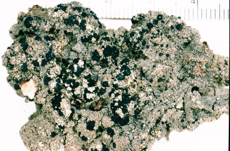
Trapeliopsis granulosa (Hoffm.) Lumbsch, 1983
Description: Photograph of a herbarium specimen, now in the Lichen Herbarium of the New York Botanical Garden.Copyright: Ed Uebel
License: CC BY-SA 3.0
Identified by: Richard E. Riefner
Date: 1981-06-27 00:00:00

Trapeliopsis granulosa (Hoffm.) Lumbsch, 1983
Description: Photograph of a herbarium specimen, now at the Lichen Herbarium of the New York Botanical Garden (scale marks = millimeters).Copyright: Ed Uebel
License: CC BY-SA 3.0
Identified by: Richard E. Riefner
Date: 1981-06-27 00:00:00

Trapeliopsis granulosa (Hoffm.) Lumbsch, 1983
Description: Photograph of a herbarium specimen, now at the Lichen Herbarium of the New York Botanical Garden (scale marks = millimeters).Copyright: Ed Uebel
License: CC BY-SA 3.0
Identified by: Richard E. Riefner
Date: 1981-06-27 00:00:00

Trapeliopsis granulosa (Hoffm.) Lumbsch, 1983
Description: Photograph of apothecia through a dissecting microscope, x30. Specimen is now in the Lichen Herbarium of the New York Botanical Garden.Copyright: Ed Uebel
License: CC BY-SA 3.0
Identified by: Richard E. Riefner
Date: 1981-06-27 00:00:00
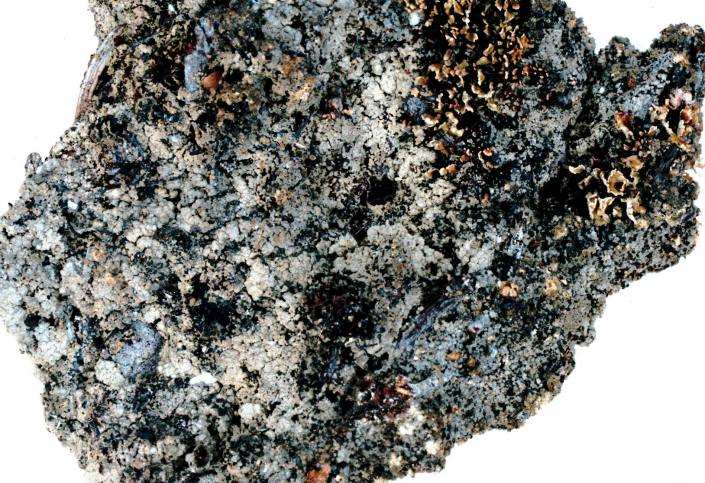
Trapeliopsis granulosa (Hoffm.) Lumbsch, 1983
Description: Photograph of a herbarium specimen showing a thallus of granular to hemispherical verrucae with some verrucae breaking down to coarse soredia; apothecia dark brown or blackish.Copyright: Ed Uebel
License: CC BY-SA 3.0
Identified by: David H. S. Richardson
Date: 2005-05-02 00:00:00

Trapeliopsis granulosa (Hoffm.) Lumbsch, 1983
Description: Photograph of a herbarium specimen showing a thallus of granular to hemispherical verrucae with some verrucae breaking down to coarse soredia; apothecia dark brown or blackish.Copyright: Ed Uebel
License: CC BY-SA 3.0
Identified by: David H. S. Richardson
Date: 2005-05-02 00:00:00
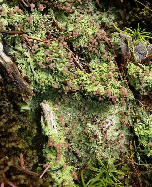
Baeomyces rufus (Huds.) Rebent.
Copyright: Bernd HaynoldLicense: CC BY-SA 3.0
Date: 2008-02-28 00:00:00
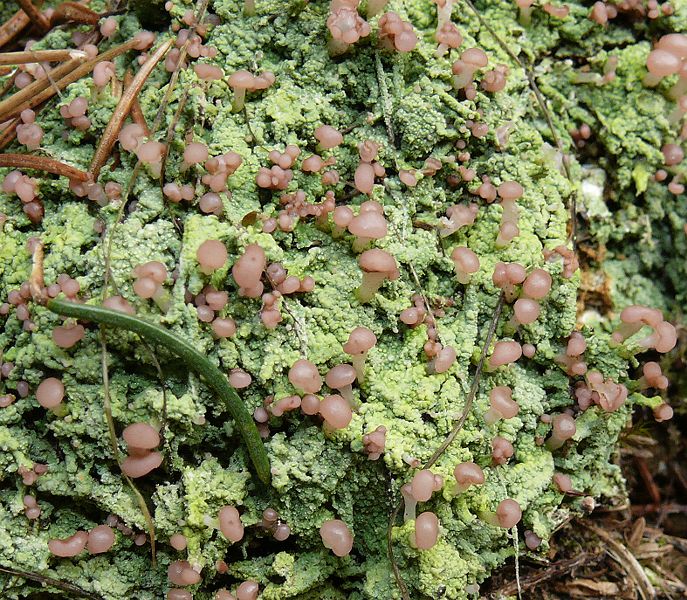
Baeomyces rufus (Huds.) Rebent.
Copyright: Bernd HaynoldLicense: CC BY-SA 3.0
Date: 2008-02-28 00:00:00

Candelaria concolor (Dicks.) Stein, 1879
Copyright: Ed UebelLicense: All Rights Reserved
Date: 0000-00-00 00:00:00

Candelaria concolor (Dicks.) Stein, 1879
Description: Photograph of a herbarium specimen taken through a dissecting microscope (x8) showing small yellow lobes edged with yellow granular sorediaCopyright: Ed Uebel
License: CC BY-SA 3.0
Identified by: Elmer G. Worthley
Date: 0000-00-00 00:00:00

Candelaria concolor (Dicks.) Stein, 1879
Copyright: Ed UebelLicense: All Rights Reserved
Date: 0000-00-00 00:00:00

Candelaria concolor (Dicks.) Stein, 1879
Description: Photograph of a herbarium specimen taken through a dissecting microscope (x40) showing small yellow lobes edged with yellow, granular sorediaCopyright: Ed Uebel
License: CC BY-SA 3.0
Identified by: Elmer G. Worthley
Date: 1983-04-03 00:00:00

Candelaria concolor (Dicks.) Stein, 1879
Copyright: Ed UebelLicense: All Rights Reserved
Date: 0000-00-00 00:00:00
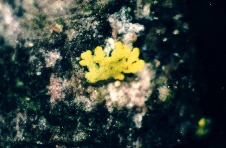
Candelaria concolor (Dicks.) Stein, 1879
Copyright: Ed UebelLicense: All Rights Reserved
Date: 0000-00-00 00:00:00

Candelaria concolor (Dicks.) Stein, 1879
Copyright: Ed UebelLicense: CC BY-SA 3.0
Date: 2003-05-08 00:00:00

Candelariella vitellina (Ehrh.) Müll. Arg., 1894
Copyright: Ed UebelLicense: All Rights Reserved
Date: 0000-00-00 00:00:00

Candelariella vitellina (Ehrh.) Müll. Arg., 1894
Copyright: Ed UebelLicense: All Rights Reserved
Date: 0000-00-00 00:00:00

Candelariella vitellina (Ehrh.) Müll. Arg., 1894
Description: Photograph of a herbarium specimen taken through a dissecting microscope (x40) showing cushions of flattened granules with apotheciaCopyright: Ed Uebel
License: CC BY-SA 3.0
Identified by: A. Norden & B. Norden
Date: 1973-08-27 00:00:00

Candelariella vitellina (Ehrh.) Müll. Arg., 1894
Copyright: Ed UebelLicense: All Rights Reserved
Date: 0000-00-00 00:00:00

Candelariella vitellina (Ehrh.) Müll. Arg., 1894
Copyright: Ed UebelLicense: All Rights Reserved
Date: 0000-00-00 00:00:00

Candelariella vitellina (Ehrh.) Müll. Arg., 1894
Copyright: Ed UebelLicense: All Rights Reserved
Date: 0000-00-00 00:00:00

Candelariella vitellina (Ehrh.) Müll. Arg., 1894
Copyright: Ed UebelLicense: All Rights Reserved
Date: 0000-00-00 00:00:00
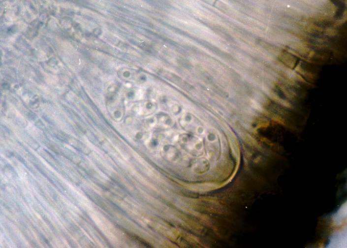
Candelariella vitellina (Ehrh.) Müll. Arg., 1894
Description: Photograph of an ascus from an apothecium of Candelariella vitellina (photographed through a compound microscope, x1000) showing numerous small spores per ascus. Commonly 16-32 spores per ascus, measuring 9-15 x 4-6.5 microns.Copyright: Ed Uebel
License: CC BY-SA 3.0
Identified by: E. C. Uebel
Date: 2006-07-19 00:00:00

Candelariella vitellina (Ehrh.) Müll. Arg., 1894
Copyright: Ed UebelLicense: All Rights Reserved
Date: 0000-00-00 00:00:00
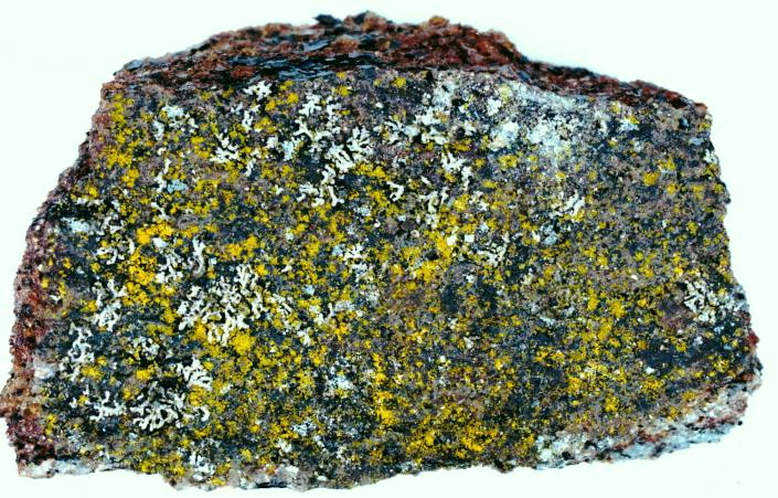
Chrysothrix candelaris (L.) J. R. Laundon, 1981
Description: Photograph of a herbarium specimen showing yellow granules. Chemical spot test: K-, yellow granules measure 30-40 microns.Copyright: Ed Uebel
License: CC BY-SA 3.0
Identified by: E. C. Uebel
Date: 2005-05-01 00:00:00
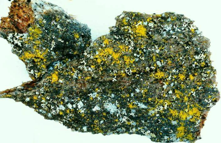
Chrysothrix candelaris (L.) J. R. Laundon, 1981
Description: Photograph of a herbarium specimen showing yellow granules. Chemical spot test: K-, yellow granules measure 30-40 microns.Copyright: Ed Uebel
License: CC BY-SA 3.0
Identified by: E. C. Uebel
Date: 2005-05-01 00:00:00

Cladonia portentosa (Dufour) Coem., 1865
Copyright: Paulo Henrique SilvaLicense: All Rights Reserved
Date: 0000-00-00 00:00:00

Cladonia portentosa (Dufour) Coem., 1865
Copyright: Paulo Henrique SilvaLicense: All Rights Reserved
Date: 0000-00-00 00:00:00

Cladonia portentosa (Dufour) Coem., 1865
Copyright: Paulo Henrique SilvaLicense: All Rights Reserved
Date: 0000-00-00 00:00:00
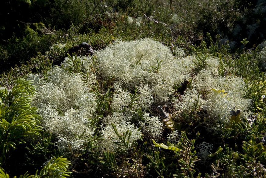
Cladonia portentosa (Dufour) Coem., 1865
Copyright: Paulo Henrique SilvaLicense: All Rights Reserved
Date: 0000-00-00 00:00:00
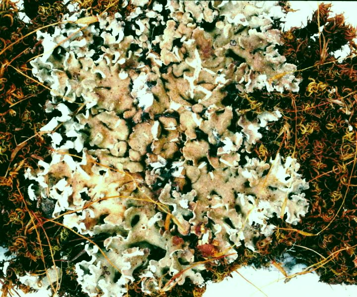
Cladonia caespiticia (Pers.) Flörke
Description: Photograph of a herbarium specimen, now in the Lichen Herbarium of the New York Botanical Garden.Copyright: Ed Uebel
License: CC BY-SA 3.0
Identified by: Richard E. Riefner
Date: 1981-07-07 00:00:00

Cladonia caespiticia (Pers.) Flörke
Copyright: James K. LindseyLicense: CC BY-SA 3.0
Date: 2005-11-22 00:00:00

Cladonia caespiticia (Pers.) Flörke
Description: Photograph of a herbarium specimen showing much divided primary squamules.Copyright: Ed Uebel
License: CC BY-SA 3.0
Identified by: E. C. Uebel
Date: 2003-06-28 00:00:00
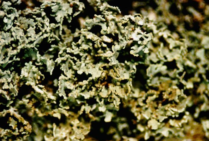
Cladonia caespiticia (Pers.) Flörke
Copyright: Ed UebelLicense: All Rights Reserved
Date: 0000-00-00 00:00:00

Cladonia caespiticia (Pers.) Flörke
Copyright: James K. LindseyLicense: All Rights Reserved
Date: 0000-00-00 00:00:00

Cladonia caespiticia (Pers.) Flörke
Description: Photograph of a herbarium specimen taken through a dissecting microscope (x30) showing several apothecia on the squamules.Copyright: Ed Uebel
License: CC BY-SA 3.0
Identified by: E. C. Uebel
Date: 2003-06-28 00:00:00

Cladonia caespiticia (Pers.) Flörke
Copyright: Ed UebelLicense: CC BY-SA 3.0
Date: 2006-08-30 00:00:00

Cladonia caespiticia (Pers.) Flörke
Copyright: Ed UebelLicense: All Rights Reserved
Date: 0000-00-00 00:00:00

Cladonia caespiticia (Pers.) Flörke
Description: Photograph of a herbarium specimen showing brown apothecia at the tips of podetia.Copyright: Ed Uebel
License: CC BY-SA 3.0
Identified by: David H. S. Richardson
Date: 2005-05-02 00:00:00

Cladonia cariosa (Ach.) Spreng.
Description: Photograph of a herbarium specimen taken through a dissecting microscope (x21) showing chocolate-brown apothecia, specimen is now in the Lichen Herbarium of the New York Botanical Garden.Copyright: Ed Uebel
License: CC BY-SA 3.0
Identified by: Elmer G. Worthley
Date: 1980-03-09 00:00:00

Cladonia cariosa (Ach.) Spreng.
Description: Photograph of a herbarium specimen, now in the Lichen Herbarium of the New York Botanical Garden.Copyright: Ed Uebel
License: CC BY-SA 3.0
Identified by: Richard E. Riefner
Date: 1981-07-14 00:00:00

Cladonia cariosa (Ach.) Spreng.
Description: Photograph of a herbarium specimen taken through a dissecting microscope (x15) showing chocolate-brown apothecia, specimen is now in the Lichen Herbarium of the New York Botanical Garden.Copyright: Ed Uebel
License: CC BY-SA 3.0
Identified by: Richard E. Riefner
Date: 1981-07-14 00:00:00

Cladonia cariosa (Ach.) Spreng.
Description: Photograph of a herbarium specimen taken through a dissecting microscope (x30) showing chocolate-brown apothecia, specimen is now in the Lichen Herbarium of the New York Botanical Garden.Copyright: Ed Uebel
License: CC BY-SA 3.0
Identified by: Richard E. Riefner
Date: 1981-07-14 00:00:00

Cladonia cariosa (Ach.) Spreng.
Description: Photograph of a herbarium specimen, now in the Lichen Herbarium of the New York Botanical Garden.Copyright: Ed Uebel
License: CC BY-SA 3.0
Identified by: Elmer G. Worthley
Date: 1980-03-09 00:00:00

Cladonia cariosa (Ach.) Spreng.
Description: Photograph of a herbarium specimen, now in the Lichen Herbarium of the New York Botanical Garden.Copyright: Ed Uebel
License: CC BY-SA 3.0
Identified by: Elmer G. Worthley
Date: 1980-03-09 00:00:00

Cladonia cervicornis (Ach.) Flot., 1849
Copyright: Ed UebelLicense: All Rights Reserved
Date: 0000-00-00 00:00:00

Cladonia cervicornis (Ach.) Flot., 1849
Description: Photograph of a herbarium specimen taken through a dissecting microscope (x6) showing two tier proliferations from the centers of cups, specimen is now in the Lichen Herbarium of the New York Botanical Garden. (millimeter scale)Copyright: Ed Uebel
License: CC BY-SA 3.0
Identified by: Elmer G. Worthley
Date: 1983-04-01 00:00:00

Cladonia cervicornis (Ach.) Flot., 1849
Copyright: Ed UebelLicense: All Rights Reserved
Date: 0000-00-00 00:00:00

Cladonia cervicornis (Ach.) Flot., 1849
Description: Photograph of a herbarium specimenCopyright: Ed Uebel
License: CC BY-SA 3.0
Identified by: E. C. Uebel
Date: 2005-05-01 00:00:00

Cladonia cervicornis (Ach.) Flot., 1849
Copyright: Ed UebelLicense: All Rights Reserved
Date: 0000-00-00 00:00:00

Cladonia cervicornis (Ach.) Flot., 1849
Copyright: Ed UebelLicense: All Rights Reserved
Date: 0000-00-00 00:00:00

Cladonia cervicornis (Ach.) Flot., 1849
Copyright: Ed UebelLicense: All Rights Reserved
Date: 0000-00-00 00:00:00

Cladonia cervicornis (Ach.) Flot., 1849
Description: Photograph of a herbarium specimen, now in the Lichen Herbarium of the New York Botanical Garden.Copyright: Ed Uebel
License: CC BY-SA 3.0
Identified by: Elmer G. Worthley
Date: 1985-09-22 00:00:00

Cladonia cervicornis (Ach.) Flot., 1849
Copyright: Ed UebelLicense: All Rights Reserved
Date: 0000-00-00 00:00:00

Cladonia cervicornis (Ach.) Flot., 1849
Copyright: Ed UebelLicense: All Rights Reserved
Date: 0000-00-00 00:00:00

Cladonia ciliata Stirt.
Copyright: James K. LindseyLicense: CC BY-SA 3.0
Date: 2004-09-02 00:00:00

Cladonia ciliata Stirt.
Copyright: James K. LindseyLicense: CC BY-SA 3.0
Date: 2004-09-02 00:00:00

Cladonia coccifera (L.) Willd.
Copyright: James K. LindseyLicense: CC BY-SA 3.0
Date: 2007-03-02 00:00:00

Cladonia coccifera (L.) Willd.
Copyright: James K. LindseyLicense: CC BY-SA 3.0
Date: 2007-03-06 00:00:00

Cladonia coniocraea (Flörke) Spreng.
Copyright: Ed UebelLicense: All Rights Reserved
Date: 0000-00-00 00:00:00

Cladonia coniocraea (Flörke) Spreng.
Copyright: Ed UebelLicense: All Rights Reserved
Date: 0000-00-00 00:00:00

Cladonia coniocraea (Flörke) Spreng.
Copyright: Ed UebelLicense: All Rights Reserved
Date: 0000-00-00 00:00:00

Cladonia coniocraea (Flörke) Spreng.
Copyright: James K. LindseyLicense: CC BY-SA 3.0
Date: 2004-08-20 00:00:00

Cladonia coniocraea (Flörke) Spreng.
Description: Photograph of a herbarium specimen taken through a dissecting microscope (x6) showing powderhorn-shaped podetia arising from the center of primary squamules, specimen is now in the Lichen Herbarium of the New York Botanical Garden.Copyright: Ed Uebel
License: CC BY-SA 3.0
Identified by: Richard E. Riefner
Date: 1981-06-23 00:00:00

Cladonia coniocraea (Flörke) Spreng.
Description: Photograph of a herbarium specimen taken through a dissecting microscope (x20) showing powderhorn-shaped podetia covered with granular soredia, specimen is now in the Lichen Herbarium of the New York Botanical Garden.Copyright: Ed Uebel
License: CC BY-SA 3.0
Identified by: Richard E. Riefner
Date: 1981-06-23 00:00:00

Cladonia coniocraea (Flörke) Spreng.
Description: Photograph of a herbarium specimen.Copyright: Ed Uebel
License: CC BY-SA 3.0
Identified by: E. C. Uebel
Date: 2005-05-02 00:00:00

Cladonia coniocraea (Flörke) Spreng.
Copyright: Ed UebelLicense: All Rights Reserved
Date: 0000-00-00 00:00:00

Cladonia coniocraea (Flörke) Spreng.
Copyright: Ed UebelLicense: All Rights Reserved
Date: 0000-00-00 00:00:00

Cladonia coniocraea (Flörke) Spreng.
Copyright: James K. LindseyLicense: CC BY-SA 3.0
Date: 2004-08-20 00:00:00

Cladonia coniocraea (Flörke) Spreng.
Copyright: James K. LindseyLicense: CC BY-SA 3.0
Date: 2007-03-02 00:00:00

Cladonia foliacea (Huds.) Willd.
Copyright: Bernd HaynoldLicense: CC BY-SA 3.0
Date: 2008-03-09 00:00:00

Cladonia foliacea (Huds.) Willd.
Copyright: Bernd HaynoldLicense: CC BY-SA 3.0
Date: 2007-10-28 00:00:00

Cladonia foliacea (Huds.) Willd.
Copyright: ZirpeLicense: CC BY-SA 3.0
Date: 2006-10-29 00:00:00
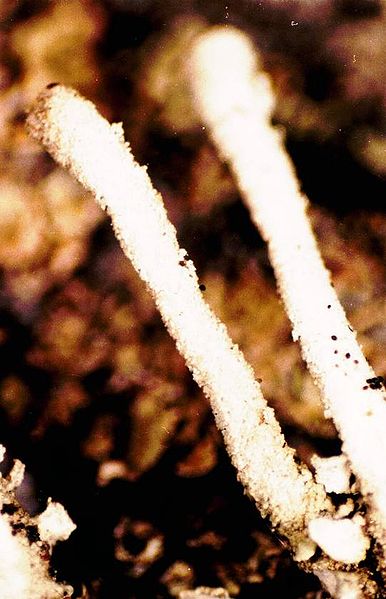
Cladonia floerkeana (Fr.) Flörke
Copyright: Ed UebelLicense: All Rights Reserved
Date: 0000-00-00 00:00:00

Cladonia floerkeana (Fr.) Flörke
Description: Photograph of a herbarium specimen taken through a dissecting microscope (x12) showing podetia covered with coarsely granular soredia, specimen is now in the Lichen Herbarium of the New York Botanical Garden.Copyright: Ed Uebel
License: CC BY-SA 3.0
Identified by: Richard E. Riefner
Date: 1981-07-14 00:00:00

Cladonia floerkeana (Fr.) Flörke
Description: Photograph of a herbarium specimen, now in the Lichen Herbarium of the New York Botanical Garden.Copyright: Ed Uebel
License: CC BY-SA 3.0
Identified by: Richard E. Riefner
Date: 1981-07-14 00:00:00

Cladonia floerkeana (Fr.) Flörke
Copyright: Ed UebelLicense: All Rights Reserved
Date: 0000-00-00 00:00:00

Cladonia floerkeana (Fr.) Flörke
Copyright: James K. LindseyLicense: CC BY-SA 3.0
Date: 2004-09-02 00:00:00

Cladonia furcata (Huds.) Schrad.
Copyright: Ed UebelLicense: All Rights Reserved
Date: 0000-00-00 00:00:00

Cladonia furcata (Huds.) Schrad.
Description: Photograph of a herbarium specimen. Note the squamules on the podetium.Copyright: Ed Uebel
License: CC BY-SA 3.0
Identified by: David H. S. Richardson
Date: 2005-05-02 00:00:00

Cladonia furcata (Huds.) Schrad.
Description: Photograph of a herbarium specimen. Note the squamules on the podetium.Copyright: Ed Uebel
License: CC BY-SA 3.0
Identified by: Don G. Flenniken
Date: 2005-05-02 00:00:00

Cladonia furcata (Huds.) Schrad.
Description: Photograph of a herbarium specimen, now in the Lichen Herbarium of the New York Botanical Garden.Copyright: Ed Uebel
License: CC BY-SA 3.0
Identified by: A. Norden & B. Ball
Date: 1972-12-31 00:00:00

Cladonia furcata (Huds.) Schrad.
Copyright: James K. LindseyLicense: CC BY-SA 3.0
Date: 2004-10-25 00:00:00

Cladonia furcata (Huds.) Schrad.
Description: Photograph of a herbarium specimen; note the podetial squamules. Specimen is now in the Lichen Herbarium of the New York Botanical Garden.Copyright: Ed Uebel
License: CC BY-SA 3.0
Date: 1972-12-31 00:00:00

Cladonia furcata (Huds.) Schrad.
Copyright: Ed UebelLicense: All Rights Reserved
Date: 0000-00-00 00:00:00

Cladonia furcata (Huds.) Schrad.
Description: Photograph of a herbarium specimen, now in the Lichen Herbarium of the New York Botanical Garden.Copyright: Ed Uebel
License: CC BY-SA 3.0
Identified by: Elmer G. Worthley
Date: 1984-10-13 00:00:00

Cladonia furcata (Huds.) Schrad.
Description: Photograph of a herbarium specimen taken through a dissecting microscope (x12) showing the upper portions of podetia, specimen is now in the Lichen Herbarium of the New York Botanical Garden.Copyright: Ed Uebel
License: CC BY-SA 3.0
Identified by: Elmer G. Worthley
Date: 1984-10-13 00:00:00

Cladonia furcata (Huds.) Schrad.
Description: Photograph of a herbarium specimen taken through a dissecting microscope (x25) showing an open axil in a podetium, specimen is now in the Lichen Herbarium of the New York Botanical Garden.Copyright: Ed Uebel
License: CC BY-SA 3.0
Identified by: Elmer G. Worthley
Date: 1984-10-13 00:00:00

Cladonia furcata (Huds.) Schrad.
Description: Photograph of a herbarium specimen taken through a dissecting microscope (x40) showing an open axil in a podetium, specimen is now in the Lichen Herbarium of the New York Botanical Garden.Copyright: Ed Uebel
License: CC BY-SA 3.0
Identified by: Elmer G. Worthley
Date: 1984-10-13 00:00:00

Cladonia furcata (Huds.) Schrad.
Copyright: James K. LindseyLicense: CC BY-SA 3.0
Date: 2004-10-25 00:00:00

Cladonia gracilis (L.) Willd.
Description: Photograph of herbarium specimens showing cup-bearing podetia with a smooth, unbroken cortex, and no soredia.Copyright: Ed Uebel
License: CC BY-SA 3.0
Identified by: David H. S. Richardson
Date: 2005-05-02 00:00:00

Cladonia gracilis (L.) Willd.
Copyright: James K. LindseyLicense: CC BY-SA 3.0
Date: 2002-11-27 00:00:00

Cladonia gracilis (L.) Willd.
Copyright: James K. LindseyLicense: CC BY-SA 3.0
Date: 2002-11-27 00:00:00

Cladonia macilenta Hoffm.
Copyright: James K. LindseyLicense: CC BY-SA 3.0
Date: 2004-09-02 00:00:00

Cladonia macilenta Hoffm.
Description: Photograph of a herbarium specimen. (Spot test reactions on podetia: PD+ dark yellow, K+ yellow)Copyright: Ed Uebel
License: CC BY-SA 3.0
Identified by: E. C. Uebel
Date: 2005-05-02 00:00:00

Cladonia peziziformis (With.) J. R. Laundon
Copyright: Ed UebelLicense: CC BY-SA 3.0
Date: 2006-08-29 00:00:00

Cladonia peziziformis (With.) J. R. Laundon
Description: Photograph of a herbarium specimen, now in the Lichen Herbarium of the New York Botanical Garden (millimeter scale).Copyright: Ed Uebel
License: CC BY-SA 3.0
Identified by: Elmer G. Worthley
Date: 1983-04-03 00:00:00

Cladonia peziziformis (With.) J. R. Laundon
Description: Photograph of a herbarium specimen showing small, rounded primary squamules, specimen is now in the Lichen Herbarium of the New York Botanical Garden.Copyright: Ed Uebel
License: CC BY-SA 3.0
Identified by: Elmer G. Worthley
Date: 1983-04-03 00:00:00

Cladonia peziziformis (With.) J. R. Laundon
Description: Photograph of a herbarium specimen taken through a dissecting microscope (x10) showing podetia topped with large, pale brown, turban-like apothecia, specimen is now in the Lichen Herbarium of the New York Botanical Garden.Copyright: Ed Uebel
License: CC BY-SA 3.0
Identified by: Elmer G. Worthley
Date: 1983-04-03 00:00:00

Cladonia peziziformis (With.) J. R. Laundon
Description: Photograph of a herbarium specimen.Copyright: Ed Uebel
License: CC BY-SA 3.0
Identified by: E. C. Uebel
Date: 2005-05-02 00:00:00

Cladonia peziziformis (With.) J. R. Laundon
Description: Photograph of a herbarium specimen.Copyright: Ed Uebel
License: CC BY-SA 3.0
Identified by: E. C. Uebel
Date: 2005-05-02 00:00:00

Cladonia peziziformis (With.) J. R. Laundon
Description: Photograph of a herbarium specimen.Copyright: Ed Uebel
License: CC BY-SA 3.0
Identified by: E. C. Uebel
Date: 2005-05-02 00:00:00

Cladonia peziziformis (With.) J. R. Laundon
Description: Photographed in the field.Copyright: Ed Uebel
License: CC BY-SA 3.0
Identified by: E. C. Uebel
Date: 2003-05-09 00:00:00

Cladonia peziziformis (With.) J. R. Laundon
Description: Photographed in the field.Copyright: Ed Uebel
License: CC BY-SA 3.0
Identified by: E. C. Uebel
Date: 2003-05-09 00:00:00

Cladonia peziziformis (With.) J. R. Laundon
Description: Photographed in the field.Copyright: Ed Uebel
License: CC BY-SA 3.0
Identified by: E. C. Uebel
Date: 2003-05-09 00:00:00

Cladonia peziziformis (With.) J. R. Laundon
Description: Photographed in the field.Copyright: Ed Uebel
License: CC BY-SA 3.0
Identified by: E. C. Uebel
Date: 2003-05-09 00:00:00

Cladonia peziziformis (With.) J. R. Laundon
Description: Photographed in the field. Photo shows the large, pale brown, turban-like apothecia of C. peziziformis.Copyright: Ed Uebel
License: CC BY-SA 3.0
Identified by: E. C. Uebel
Date: 2001-09-15 00:00:00

Cladonia pyxidata (L.) Hoffm.
Copyright: Kristian PetersLicense: CC BY-SA 3.0
Date: 2006-12-10 00:00:00

Cladonia pyxidata (L.) Hoffm.
Description: Photograph of a herbarium specimen, now in the Lichen Herbarium of the New York Botanical Garden (millimeter scale).Copyright: Ed Uebel
License: CC BY-SA 3.0
Identified by: Elmer G. Worthley
Date: 1984-02-16 00:00:00

Cladonia pyxidata (L.) Hoffm.
Description: Photograph of a herbarium specimen taken through a dissecting microscope (x40) showing round areoles inside the cup, specimen is now in the Lichen Herbarium of the New York Botanical Garden.Copyright: Ed Uebel
License: CC BY-SA 3.0
Identified by: Elmer G. Worthley
Date: 1984-02-16 00:00:00

Cladonia pyxidata (L.) Hoffm.
Description: Photograph of a herbarium specimen taken through a dissecting microscope (x15) showing flat areoles inside the cup, specimen is now in the Lichen Herbarium of the New York Botanical Garden.Copyright: Ed Uebel
License: CC BY-SA 3.0
Identified by: Elmer G. Worthley
Date: 1984-02-16 00:00:00

Cladonia pyxidata (L.) Hoffm.
Description: Photograph of a herbarium specimen, now in the Lichen Herbarium of the New York Botanical Garden (millimeter scale).Copyright: Ed Uebel
License: CC BY-SA 3.0
Identified by: Elmer G. Worthley & Allen C. Skorepa
Date: 1984-08-26 00:00:00

Cladonia pyxidata (L.) Hoffm.
Description: Photograph of a herbarium specimen taken through a dissecting microscope (x10) showing cups with areoles on the inner surface, and brown proliferating apothecia), specimen is now in the Lichen Herbarium of the New York Botanical Garden.Copyright: Ed Uebel
License: CC BY-SA 3.0
Identified by: Elmer G. Worthley & Allen C. Skorepa
Date: 1984-08-26 00:00:00

Cladonia pyxidata (L.) Hoffm.
Description: Photograph of a herbarium specimenCopyright: Ed Uebel
License: CC BY-SA 3.0
Identified by: E. C. Uebel
Date: 2005-05-02 00:00:00

Cladonia pyxidata (L.) Hoffm.
Description: Photograph of a herbarium specimenCopyright: Ed Uebel
License: CC BY-SA 3.0
Identified by: E. C. Uebel
Date: 2005-05-02 00:00:00

Cladonia pyxidata (L.) Hoffm.
Copyright: Kristian PetersLicense: CC BY-SA 3.0
Date: 2006-11-08 00:00:00

Cladonia pyxidata (L.) Hoffm.
Copyright: OhWehLicense: CC-BY-SA-2.5
Date: 2008-05-03 00:00:00

Cladonia pyxidata (L.) Hoffm.
Copyright: OhWehLicense: CC-BY-SA-2.5
Date: 2008-05-03 00:00:00

Cladonia pyxidata (L.) Hoffm.
Copyright: OhWehLicense: CC-BY-SA-2.5
Date: 2008-05-03 00:00:00

Cladonia squamosa (Scop.) Hoffm., 1796
Copyright: Ed UebelLicense: All Rights Reserved
Date: 0000-00-00 00:00:00

Cladonia squamosa (Scop.) Hoffm., 1796
Copyright: Ed UebelLicense: All Rights Reserved
Date: 0000-00-00 00:00:00

Cladonia squamosa (Scop.) Hoffm., 1796
Copyright: Ed UebelLicense: All Rights Reserved
Date: 0000-00-00 00:00:00

Cladonia squamosa (Scop.) Hoffm., 1796
Description: Photograph of a herbarium specimen.Copyright: Ed Uebel
License: CC BY-SA 3.0
Identified by: E. C. Uebel
Date: 2005-05-03 00:00:00

Cladonia squamosa (Scop.) Hoffm., 1796
Description: Photograph of a herbarium specimen taken through a dissecting microscope (x37) showing small cups at the tips of podetia, specimen is now in the Lichen Herbarium of the New York Botanical Garden.Copyright: Ed Uebel
License: CC BY-SA 3.0
Identified by: Elmer G. Worthley
Date: 1983-10-22 00:00:00

Cladonia squamosa (Scop.) Hoffm., 1796
Description: Photograph of a herbarium specimen, now in the Lichen Herbarium of the New York Botanical Garden (millimeter scale).Copyright: Ed Uebel
License: CC BY-SA 3.0
Identified by: A. Norden & B. Ball
Date: 1973-03-04 00:00:00

Cladonia squamosa (Scop.) Hoffm., 1796
Copyright: James K. LindseyLicense: CC BY-SA 3.0
Date: 2006-09-09 00:00:00

Cladonia squamosa (Scop.) Hoffm., 1796
Copyright: Ed UebelLicense: All Rights Reserved
Date: 0000-00-00 00:00:00

Cladonia squamosa (Scop.) Hoffm., 1796
Description: Photograph of a herbarium specimen, now in the Lichen Herbarium of the New York Botanical Garden (millimeter scale).Copyright: Ed Uebel
License: CC BY-SA 3.0
Identified by: Elmer G. Worthley
Date: 1983-10-22 00:00:00

Cladonia squamosa (Scop.) Hoffm., 1796
Description: Photograph of a herbarium specimen taken through a dissecting microscope (x6) showing squamulose podetia, specimen is now in the Lichen Herbarium of the New York Botanical Garden.Copyright: Ed Uebel
License: CC BY-SA 3.0
Identified by: Elmer G. Worthley
Date: 1983-10-22 00:00:00

Cladonia squamosa (Scop.) Hoffm., 1796
Description: Photograph of a herbarium specimenCopyright: Ed Uebel
License: CC BY-SA 3.0
Identified by: E. C. Uebel
Date: 2005-05-03 00:00:00

Cladonia squamosa (Scop.) Hoffm., 1796
Copyright: James K. LindseyLicense: CC BY-SA 3.0
Date: 2006-09-09 00:00:00

Coenogonium luteum (Dicks.) Kalb & Lücking
Description: Photograph of a herbarium specimen, now in the Lichen Herbarium of the New York Botanical Garden (millimeter scale).Copyright: Ed Uebel
License: CC BY-SA 3.0
Identified by: Richard E. Riefner
Date: 1981-06-29 00:00:00

Coenogonium luteum (Dicks.) Kalb & Lücking
Description: Photograph of a herbarium specimen taken through a dissecting microscope (x9) showing apothecia with orange disks and pale margins, specimen is now in the Lichen Herbarium of the New York Botanical Garden.Copyright: Ed Uebel
License: CC BY-SA 3.0
Identified by: Richard E. Riefner
Date: 1981-06-29 00:00:00

Coenogonium luteum (Dicks.) Kalb & Lücking
Description: Photograph of a herbarium specimen taken through a dissecting microscope (x34) showing apothecia with orange disks and pale margins, specimen is now in the Lichen Herbarium of the New York Botanical Garden.Copyright: Ed Uebel
License: CC BY-SA 3.0
Identified by: Richard E. Riefner
Date: 1981-06-29 00:00:00
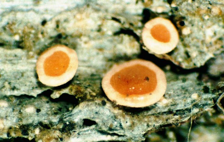
Coenogonium luteum (Dicks.) Kalb & Lücking
Description: Photograph of a herbarium specimen taken through a dissecting microscope (x40) showing apothecia with orange disks and pale margins, specimen is now in the Lichen Herbarium of the New York Botanical Garden.Copyright: Ed Uebel
License: CC BY-SA 3.0
Identified by: Richard E. Riefner
Date: 1981-06-29 00:00:00

Coenogonium pineti (Schrad.) Lücking & Lumbsch
Copyright: Ed UebelLicense: All Rights Reserved
Date: 0000-00-00 00:00:00

Coenogonium pineti (Schrad.) Lücking & Lumbsch
Copyright: Ed UebelLicense: All Rights Reserved
Date: 0000-00-00 00:00:00
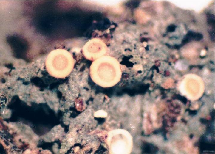
Coenogonium pineti (Schrad.) Lücking & Lumbsch
Copyright: Ed UebelLicense: All Rights Reserved
Date: 0000-00-00 00:00:00

Coenogonium pineti (Schrad.) Lücking & Lumbsch
Copyright: Ed UebelLicense: All Rights Reserved
Date: 0000-00-00 00:00:00

Collema furfuraceum Du Rietz
Description: Photograph of a herbarium specimen showing broad lobes with conspicuous ridges and pustules, specimen is now in the Lichen Herbarium of the New York Botanical Garden. (millimeter scale)Copyright: Ed Uebel
License: CC BY-SA 3.0
Identified by: A. Norden & B. Ball
Date: 1973-08-14 00:00:00

Collema furfuraceum Du Rietz
Description: Photograph of a herbarium specimen showing broad lobes with conspicuous ridges and pustules, specimen is now in the Lichen Herbarium of the New York Botanical Garden.Copyright: Ed Uebel
License: CC BY-SA 3.0
Identified by: A. Norden & B. Ball
Date: 1973-08-14 00:00:00

Collema furfuraceum Du Rietz
Description: Photograph of a herbarium specimen showing broad lobes with conspicuous ridges and pustules, specimen is now in the Lichen Herbarium of the New York Botanical Garden. (millimeter scale)Copyright: Ed Uebel
License: CC BY-SA 3.0
Identified by: A. Norden & B. Ball
Date: 1973-08-14 00:00:00

Collema furfuraceum Du Rietz
Description: Photograph of a herbarium specimen showing broad lobes with conspicuous ridges and pustules, specimen is now in the Lichen Herbarium of the New York Botanical Garden.Copyright: Ed Uebel
License: CC BY-SA 3.0
Identified by: A. Norden & B. Ball
Date: 1973-08-14 00:00:00

Collema furfuraceum Du Rietz
Description: Photograph of a herbarium specimen taken through a dissecting microscope (x6) showing broad lobes with conspicuous ridges and pustules, specimen is now in the Lichen Herbarium of the New York Botanical Garden.Copyright: Ed Uebel
License: CC BY-SA 3.0
Identified by: A. Norden & B. Ball
Date: 1973-08-14 00:00:00

Collema furfuraceum Du Rietz
Description: Photograph of a herbarium specimen taken through a dissecting microscope (x6) showing broad lobes with conspicuous ridges and pustules, specimen is now in the Lichen Herbarium of the New York Botanical Garden.Copyright: Ed Uebel
License: CC BY-SA 3.0
Identified by: A. Norden & B. Ball
Date: 1973-08-14 00:00:00

Collema furfuraceum Du Rietz
Description: Photograph of a herbarium specimen taken through a dissecting microscope (x6) showing broad lobes with conspicuous ridges and pustules, specimen is now in the Lichen Herbarium of the New York Botanical Garden.Copyright: Ed Uebel
License: CC BY-SA 3.0
Identified by: A. Norden & B. Ball
Date: 1973-08-14 00:00:00

Collema furfuraceum Du Rietz
Description: Photograph of a herbarium specimen taken through a dissecting microscope (x10) showing broad lobes with conspicuous ridges and pustules, specimen is now in the Lichen Herbarium of the New York Botanical Garden.Copyright: Ed Uebel
License: CC BY-SA 3.0
Identified by: A. Norden & B. Ball
Date: 1973-08-14 00:00:00

Collema nigrescens (Huds.) DC.
Description: Photograph of a herbarium specimen showing a black thallus with rounded overlapping lobes.Copyright: Ed Uebel
License: CC BY-SA 3.0
Identified by: E. C. Uebel
Date: 2005-05-01 00:00:00

Collema nigrescens (Huds.) DC.
Description: Photograph of a herbarium specimen taken through a dissecting microscope (x7) showing a black thallus with rounded overlapping lobes.Copyright: Ed Uebel
License: CC BY-SA 3.0
Identified by: E. C. Uebel
Date: 2005-12-15 00:00:00

Collema nigrescens (Huds.) DC.
Description: Photograph of the upper surface of a lobe taken through a compound microscope (x400) showing the absence of a cellular cortex.Copyright: Ed Uebel
License: CC BY-SA 3.0
Identified by: E. C. Uebel
Date: 2002-06-13 00:00:00

Collema subflaccidum Degel.
Description: Photograph of a herbarium specimen taken through a dissecting microscope (x15) showing dark olive lobes with globular isidia, specimen is now in the Lichen Herbarium of the New York Botanical Garden.Copyright: Ed Uebel
License: CC BY-SA 3.0
Identified by: Richard E. Riefner
Date: 1981-07-28 00:00:00

Collema subflaccidum Degel.
Description: Photograph of a herbarium specimen taken through a dissecting microscope (x7) showing dark olive lobes with globular isidia, specimen is now in the Lichen Herbarium of the New York Botanical Garden.Copyright: Ed Uebel
License: CC BY-SA 3.0
Identified by: Richard E. Riefner
Date: 1981-07-28 00:00:00
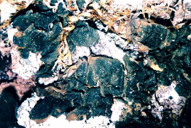
Collema subflaccidum Degel.
Description: Photograph of a herbarium specimen taken through a dissecting microscope (x6) showing dark olive lobes with globular isidia, specimen is now in the Lichen Herbarium of the New York Botanical Garden.Copyright: Ed Uebel
License: CC BY-SA 3.0
Identified by: Richard E. Riefner
Date: 1981-07-28 00:00:00

Collema subflaccidum Degel.
Description: Photograph of a herbarium specimen taken through a dissecting microscope (x40) showing globular isidia, specimen is now in the Lichen Herbarium of the New York Botanical Garden.Copyright: Ed Uebel
License: CC BY-SA 3.0
Identified by: Arnold W. Norden
Date: 1983-12-11 00:00:00

Collema subflaccidum Degel.
Description: Photograph of a herbarium specimen showing large, rounded, dark olive lobes.Copyright: Ed Uebel
License: CC BY-SA 3.0
Identified by: Don G. Flenniken
Date: 2005-05-01 00:00:00

Collema subflaccidum Degel.
Description: Photograph of a herbarium specimen showing large, rounded, dark olive lobes.Copyright: Ed Uebel
License: CC BY-SA 3.0
Identified by: Don G. Flenniken
Date: 2005-05-01 00:00:00
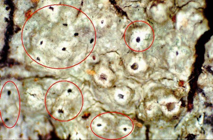
Lichenodiplis lecanorae (Vouaux) Dyko & D. Hawksw., 1979
Description: Photograph of a herbarium specimen taken through a dissecting microscope (x40) showing the black apothecia (within the red circles and ellipses) of Lichenodiplis lecanorae parasitizing the apothecia of Pertusaria xanthodes)Copyright: Ed Uebel
License: CC BY-SA 3.0
Identified by: David Hawksworth
Date: 2001-07-10 00:00:00

Lichenodiplis lecanorae (Vouaux) Dyko & D. Hawksw., 1979
Description: Photograph of a herbarium specimen taken through a dissecting microscope (x40) showing the black apothecia (within the red circles) of Lichenodiplis lecanorae parasitizing the apothecia of Pertusaria xanthodes.Copyright: Ed Uebel
License: CC BY-SA 3.0
Identified by: David Hawksworth
Date: 2001-07-10 00:00:00

Lichenodiplis lecanorae (Vouaux) Dyko & D. Hawksw., 1979
Description: Photograph of a herbarium specimen taken through a dissecting microscope (x40) showing the black apothecia of Lichenodiplis lecanorae parasitizing the apothecia of Pertusaria xanthodes. Note the two unparasitized fruiting warts with pale ostioles near theCopyright: Ed Uebel
License: CC BY-SA 3.0
Identified by: David Hawksworth
Date: 2001-07-10 00:00:00

Lichenodiplis lecanorae (Vouaux) Dyko & D. Hawksw., 1979
Description: Photograph of Lichenodiplis lecanorae spores (x1000); brown, 1-septate spores measuring approximately 6 x 3 microns. Lichenodiplis lecanorae was collected as a parasite on the apothecia of Pertusaria xanthodes.Copyright: Ed Uebel
License: CC BY-SA 3.0
Identified by: David Hawksworth
Date: 2001-07-10 00:00:00

Lichenodiplis lecanorae (Vouaux) Dyko & D. Hawksw., 1979
Description: Photograph of brown, 1-septate spores, measuring approximately 7 x 3 microns of Lichenodiplis lecanorae; magnification x1000. Lichenodiplis lecanorae was collected as a parasite on the apothecia of Pertusaria xanthodes.Copyright: Ed Uebel
License: CC BY-SA 3.0
Identified by: David Hawksworth
Date: 2001-07-10 00:00:00
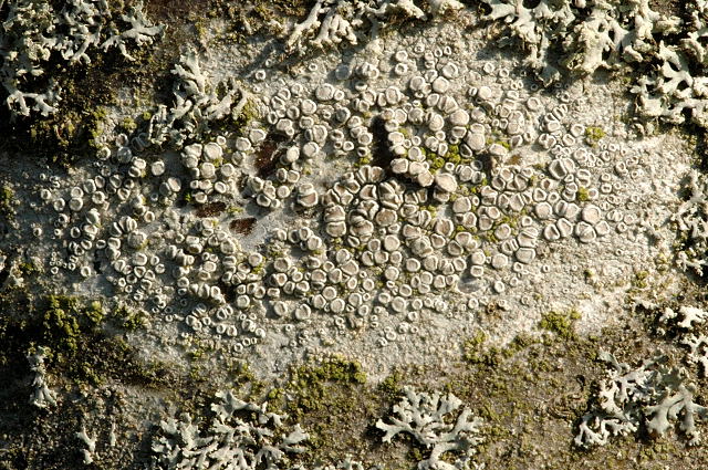
Lecanora carpinea (L.) Vain.
Copyright: James K. LindseyLicense: CC BY-SA 3.0
Date: 2004-09-03 00:00:00
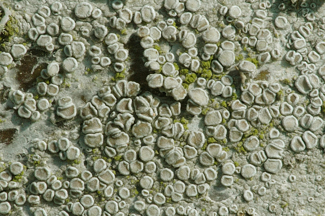
Lecanora carpinea (L.) Vain.
Copyright: James K. LindseyLicense: CC BY-SA 3.0
Date: 2004-09-04 00:00:00
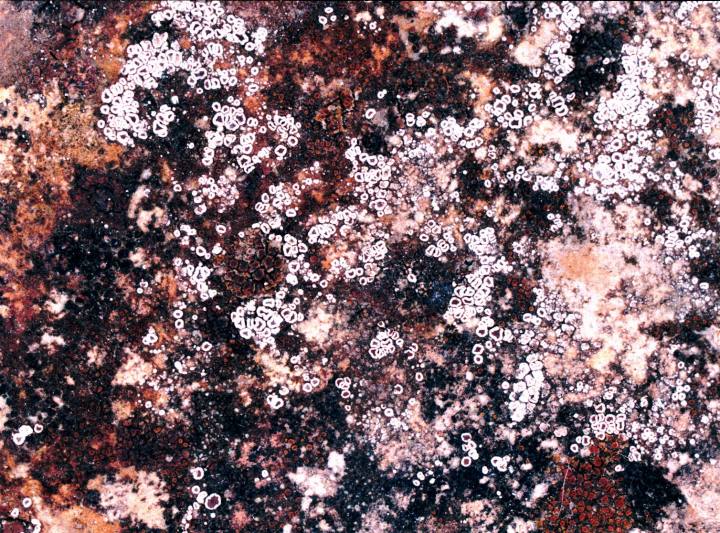
Myriolecis dispersa (Pers.) Śliwa, Zhao Xin & Lumbsch, 2015
Description: Photograph of a herbarium specimen, now in the Lichen Herbarium of the New York Botanical Garden.Copyright: Ed Uebel
License: CC BY-SA 3.0
Identified by: A. Norden & B. Norden
Date: 1974-10-29 00:00:00
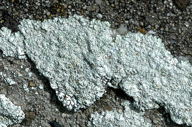
Myriolecis dispersa (Pers.) Śliwa, Zhao Xin & Lumbsch, 2015
Copyright: James K. LindseyLicense: CC BY-SA 3.0
Date: 2004-08-16 00:00:00
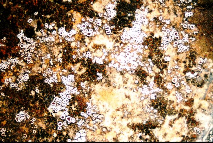
Myriolecis dispersa (Pers.) Śliwa, Zhao Xin & Lumbsch, 2015
Description: Photograph of a herbarium specimen taken through a dissecting microscope, x6, specimen is now in the Lichen Herbarium of the New York Botanical Garden.Copyright: Ed Uebel
License: CC BY-SA 3.0
Identified by: A. Norden & B. Norden
Date: 1974-10-29 00:00:00
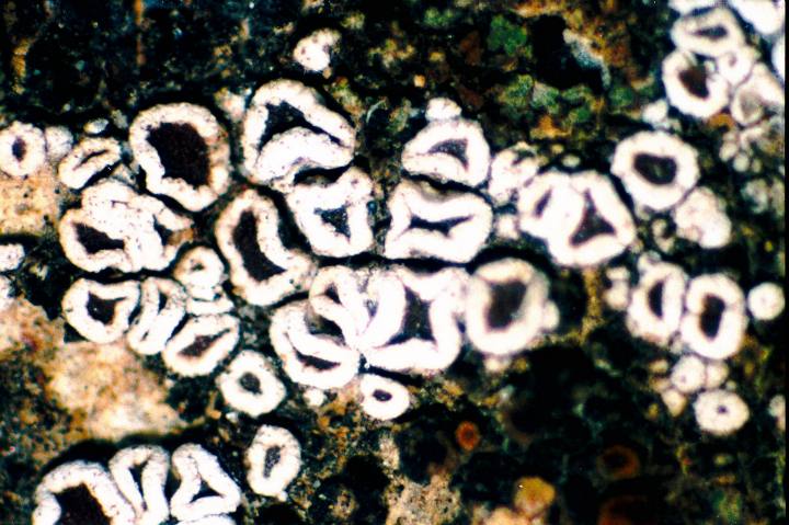
Myriolecis dispersa (Pers.) Śliwa, Zhao Xin & Lumbsch, 2015
Description: Photograph of a herbarium specimen taken through a dissecting microscope, x40 showing apothecia with brown disks and thick white margins, specimen is now in the Lichen Herbarium of the New York Botanical Garden.Copyright: Ed Uebel
License: CC BY-SA 3.0
Identified by: A. Norden & B. Norden
Date: 1974-10-29 00:00:00
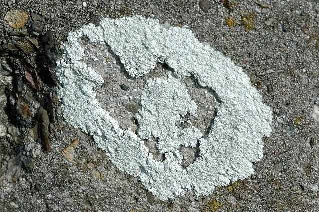
Myriolecis dispersa (Pers.) Śliwa, Zhao Xin & Lumbsch, 2015
Copyright: James K. LindseyLicense: CC BY-SA 3.0
Date: 2004-08-16 00:00:00
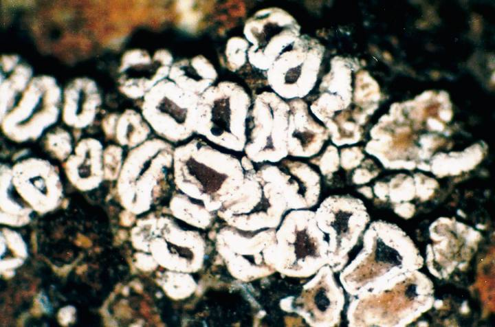
Myriolecis dispersa (Pers.) Śliwa, Zhao Xin & Lumbsch, 2015
Description: Photograph of a herbarium specimen taken through a dissecting microscope, x40 showing apothecia with brown disks and thick white margins, specimen is now in the Lichen Herbarium of the New York Botanical Garden.Copyright: Ed Uebel
License: CC BY-SA 3.0
Identified by: A. Norden & B. Norden
Date: 1974-10-29 00:00:00
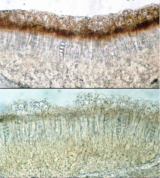
Lecanora pulicaris (Pers.) Ach.
Description: Photograph of cross sections of an apothecium from Lecanora pulicaris taken through a compound microscope (x400). The upper photo shows the epihymenium and hymenium before exposure to KOH solution. The lower photo shows the epihymenium and hymenium afterCopyright: Ed Uebel
License: CC BY-SA 3.0
Identified by: E. C. Uebel
Date: 2007-09-05 00:00:00
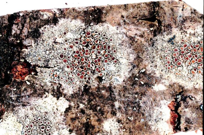
Lecanora pulicaris (Pers.) Ach.
Description: Photograph of a herbarium specimen. Spot tests on the thallus: PD+ red, K+ yellow, C-.Copyright: Ed Uebel
License: CC BY-SA 3.0
Identified by: E. C. Uebel
Date: 2007-05-01 00:00:00
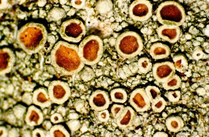
Lecanora pulicaris (Pers.) Ach.
Description: Photograph of a herbarium specimen taken through a dissecting microscope (x25), showing apothecia with reddish-brown to dark brown disks and smooth rims. Spot tests on the thallus: PD+ red, K+ yellow, C-.Copyright: Ed Uebel
License: CC BY-SA 3.0
Identified by: E. C. Uebel
Date: 2003-07-26 00:00:00
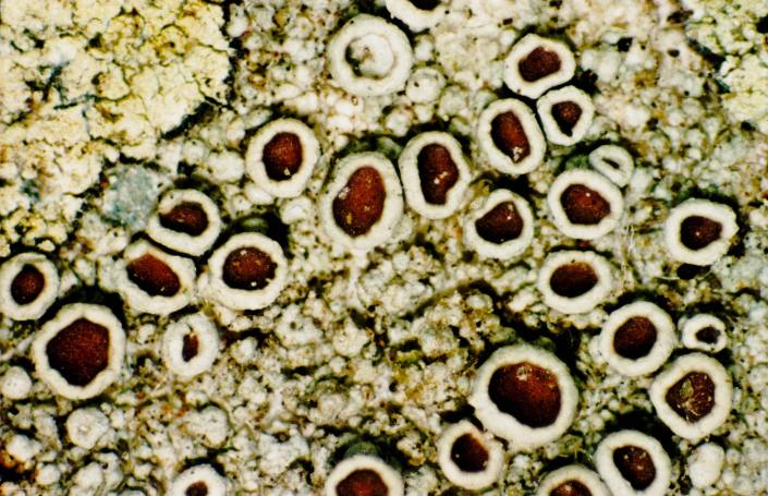
Lecanora pulicaris (Pers.) Ach.
Description: Photograph of a herbarium specimen taken through a dissecting microscope (x25), showing apothecia with reddish-brown to dark brown disks and smooth rims. Spot tests on the thallus: PD+ red, K+ yellow, C-.Copyright: Ed Uebel
License: CC BY-SA 3.0
Identified by: E. C. Uebel
Date: 2003-07-26 00:00:00
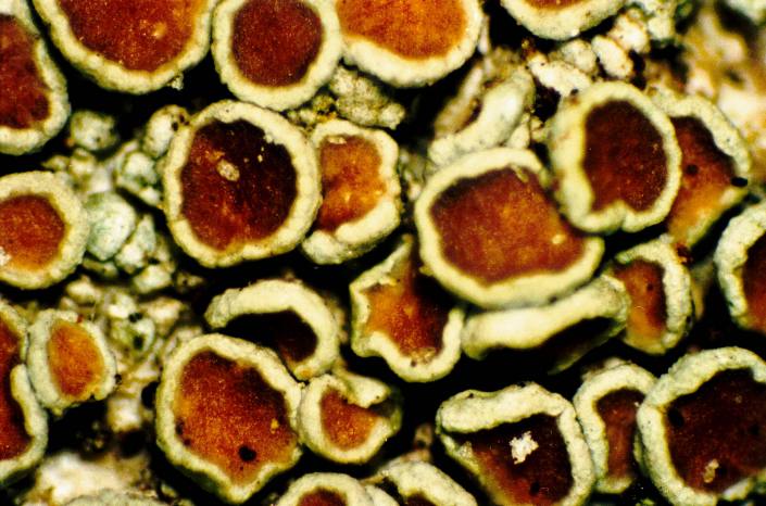
Lecanora pulicaris (Pers.) Ach.
Description: Photograph of a herbarium specimen taken through a dissecting microscope (x40), showing apothecia with reddish-brown to dark brown disks and smooth rims. Spot tests on the thallus: PD+ red, K+ yellow, C-.Copyright: Ed Uebel
License: CC BY-SA 3.0
Identified by: E. C. Uebel
Date: 2003-07-26 00:00:00
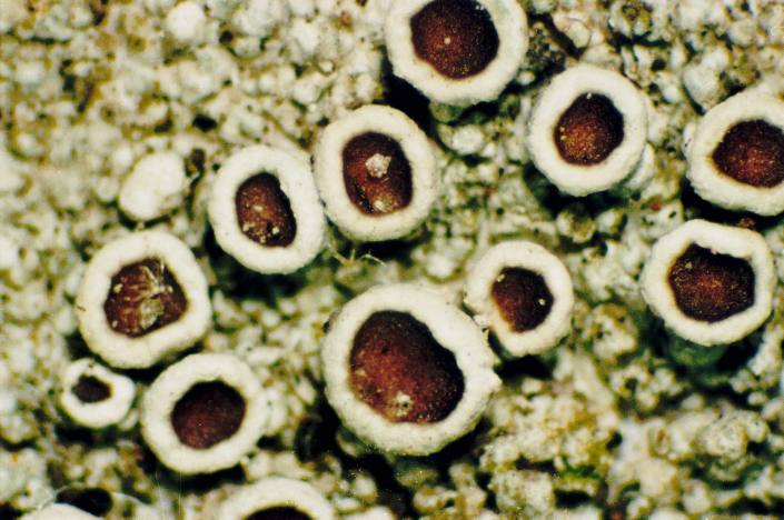
Lecanora pulicaris (Pers.) Ach.
Description: Photograph of a herbarium specimen taken through a dissecting microscope (x40), showing apothecia with reddish-brown to dark brown disks and smooth rims. Spot tests on the thallus: PD+ red, K+ yellow, C-.Copyright: Ed Uebel
License: CC BY-SA 3.0
Identified by: E. C. Uebel
Date: 2003-07-26 00:00:00
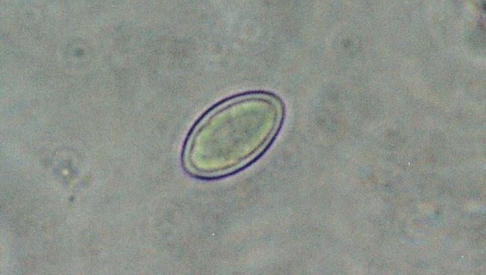
Lecanora pulicaris (Pers.) Ach.
Description: Photograph of a spore from an apothecium of Lecanora pulicaris taken through a compound microscope, x1000. (spore measures 13 x 8 microns).Copyright: Ed Uebel
License: CC BY-SA 3.0
Identified by: E. C. Uebel
Date: 2003-07-26 00:00:00
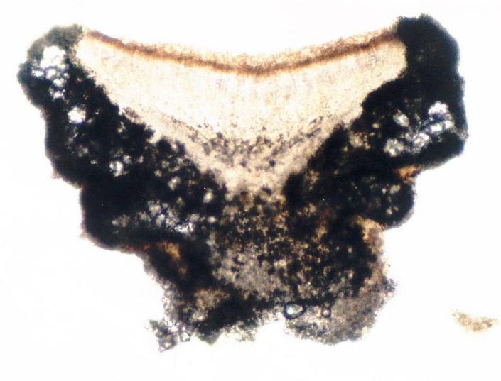
Lecanora pulicaris (Pers.) Ach.
Description: Photograph of a cross section of an apothecium from Lecanora pulicaris taken through a compound microscope (x100), showing large crystals in the amphithecium.Copyright: Ed Uebel
License: CC BY-SA 3.0
Identified by: E. C. Uebel
Date: 2007-09-05 00:00:00
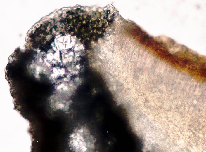
Lecanora pulicaris (Pers.) Ach.
Description: Photograph of a cross section of an apothecium from Lecanora pulicaris taken through a compound microscope (x400), showing large crystals in the amphithecium.Copyright: Ed Uebel
License: CC BY-SA 3.0
Identified by: E. C. Uebel
Date: 2007-09-05 00:00:00
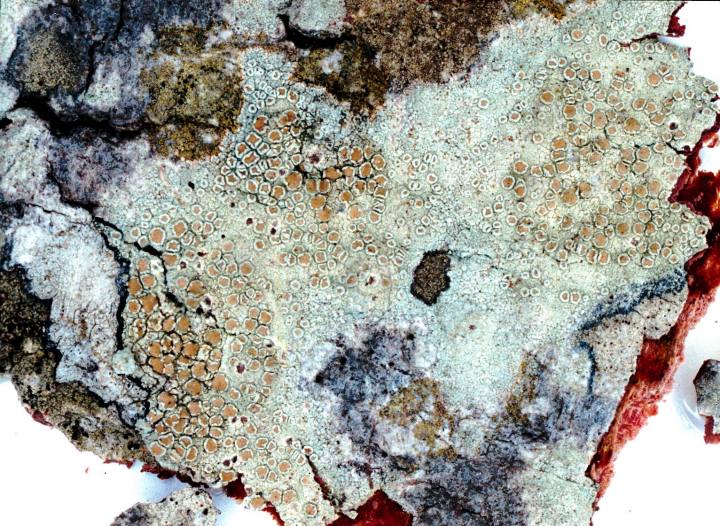
Lecanora strobilina Ach., 1814
Description: Photograph of a herbarium specimen.Copyright: Ed Uebel
License: CC BY-SA 3.0
Identified by: E. C. Uebel
Date: 2001-07-14 00:00:00
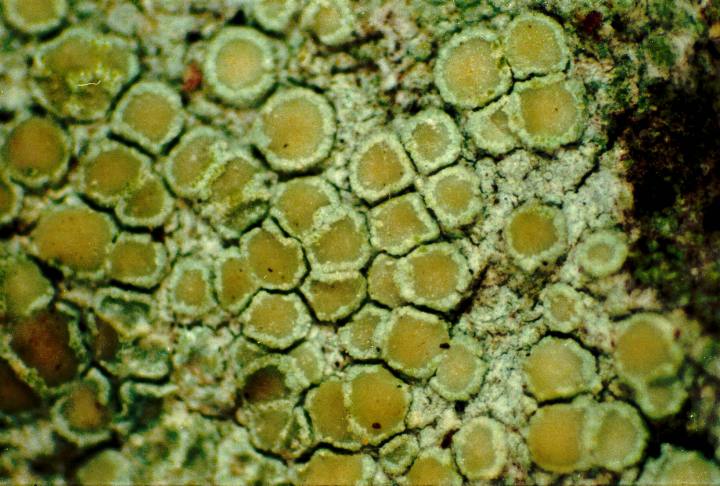
Lecanora strobilina Ach., 1814
Description: Photograph of a herbarium specimen taken through a dissecting microscope (x40) showing a pale yellowish green thallus with small, waxy light yellow apothecia. Chemical spot tests of thallus and apothecial section: PD-, K-, KC+ yellow, C- (usnic acid & zeoCopyright: Ed Uebel
License: CC BY-SA 3.0
Identified by: H. Thorsten Lumbsch
Date: 2004-10-22 00:00:00
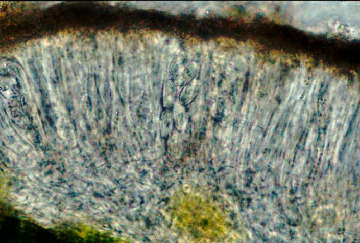
Lecanora strobilina Ach., 1814
Description: Photograph of a cross section of an apothecium, through a compound microscope (x1000), showing seven 1-celled, hyaline spores per ascus).Copyright: Ed Uebel
License: CC BY-SA 3.0
Identified by: H. Thorsten Lumbsch
Date: 2004-10-22 00:00:00
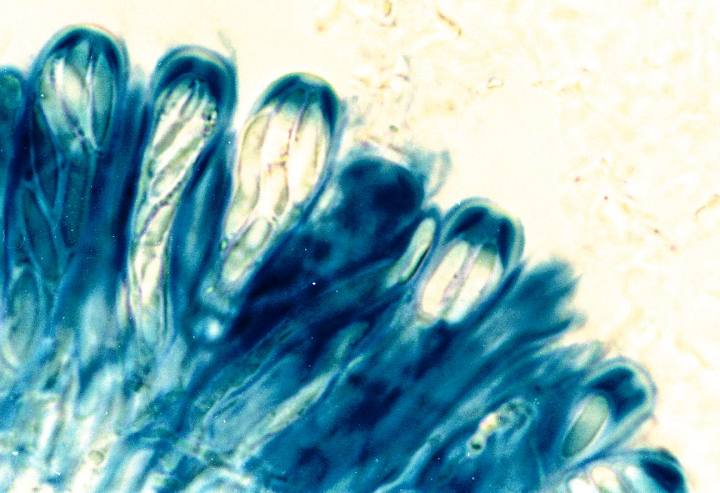
Lecanora strobilina Ach., 1814
Description: Photograph of a cross section of an apothecium, through a compound microscope (x1000), showing asci stained with iodine (Lugol's solution); note that the tips of the asci display the Lecanora-type of stainingCopyright: Ed Uebel
License: CC BY-SA 3.0
Identified by: E. C. Uebel
Date: 2005-01-01 00:00:00
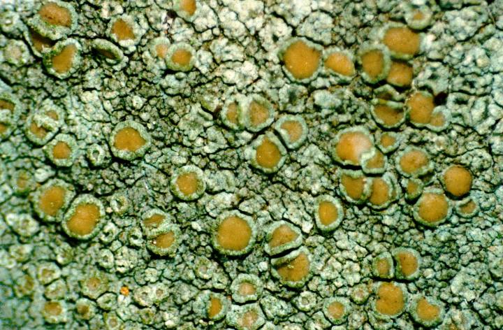
Lecanora strobilina Ach., 1814
Description: Photograph of a herbarium specimen taken through a dissecting microscope (x20) showing a pale yellowish green thallus with small, waxy light yellow apothecia. Chemical spot tests of thallus and apothecial section: PD-, K-, KC+ yellow, C- (usnic acid & zeoCopyright: Ed Uebel
License: CC BY-SA 3.0
Identified by: E. C. Uebel
Date: 2003-06-25 00:00:00
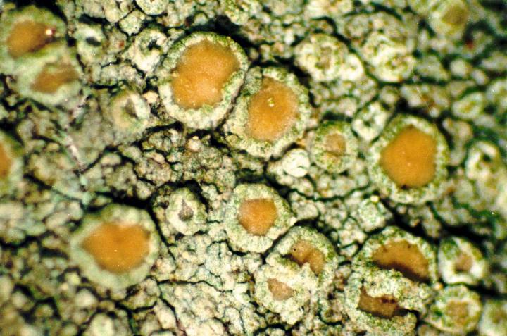
Lecanora strobilina Ach., 1814
Description: Photograph of a herbarium specimen taken through a dissecting microscope (x40) showing a pale yellowish green thallus with small, waxy light yellow apotheciaCopyright: Ed Uebel
License: CC BY-SA 3.0
Identified by: E. C. Uebel
Date: 2003-06-25 00:00:00
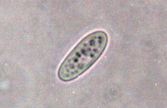
Lecanora strobilina Ach., 1814
Description: Photograph of a spore taken through a compound microscope (x1000); spore measures 12 x 5 micronsCopyright: Ed Uebel
License: CC BY-SA 3.0
Identified by: H. Thorsten Lumbsch
Date: 2004-10-22 00:00:00
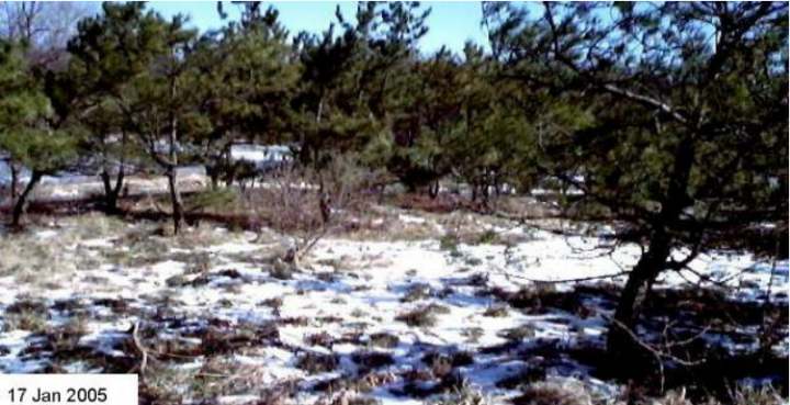
Lecanora strobilina Ach., 1814
Description: Photograph of Pinus thunbergiana trees with abundant colonies of Lecanora strobilinaCopyright: Ed Uebel
License: CC BY-SA 3.0
Date: 0000-00-00 00:00:00
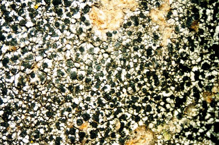
Lecidella stigmatea (Ach.) Hertel & Leuckert
Description: Photograph of a herbarium specimen taken through a dissecting microscope, x8. (Chemical spot tests on the medulla: K+ pale yellow, C-, PD-)Copyright: Ed Uebel
License: CC BY-SA 3.0
Identified by: E. C. Uebel
Date: 2006-08-01 00:00:00
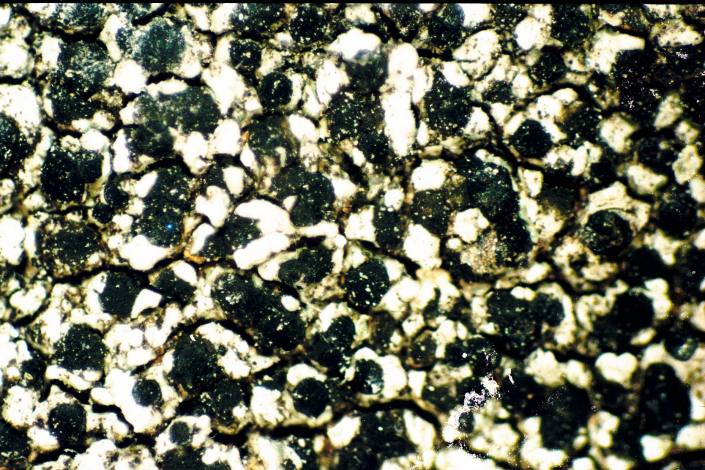
Lecidella stigmatea (Ach.) Hertel & Leuckert
Copyright: Ed UebelLicense: CC BY-SA 3.0
Date: 0000-00-00 00:00:00
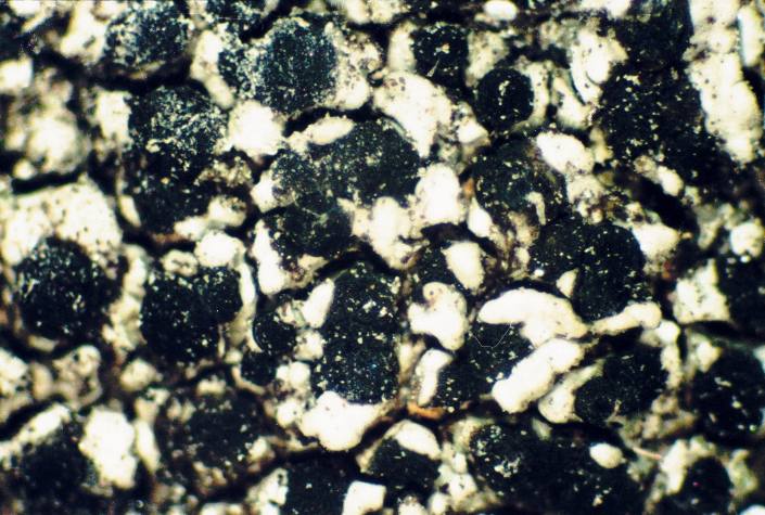
Lecidella stigmatea (Ach.) Hertel & Leuckert
Description: Photograph of a herbarium specimen taken through a dissecting microscope, x40. (Chemical spot tests on the medulla: K+ pale yellow, C-, PD-)Copyright: Ed Uebel
License: CC BY-SA 3.0
Identified by: E. C. Uebel
Date: 2006-08-01 00:00:00
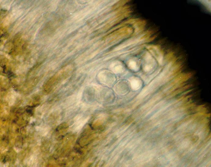
Lecidella stigmatea (Ach.) Hertel & Leuckert
Description: Photograph of an ascus from an apothecium of Lecidella stigmatea taken through a compound microscope (x1000), showing 8, simple, hyaline spores per ascus (spores measure 8 x 6 microns).Copyright: Ed Uebel
License: CC BY-SA 3.0
Identified by: E. C. Uebel
Date: 2006-08-01 00:00:00
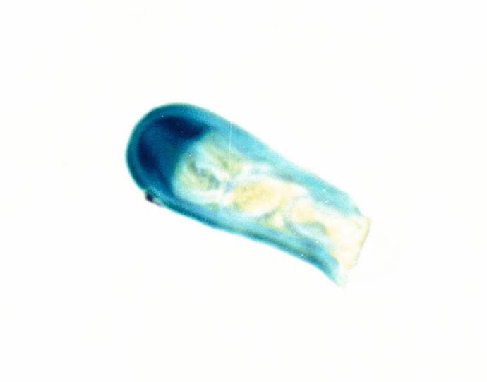
Lecidella stigmatea (Ach.) Hertel & Leuckert
Copyright: Ed UebelLicense: All Rights Reserved
Date: 0000-00-00 00:00:00
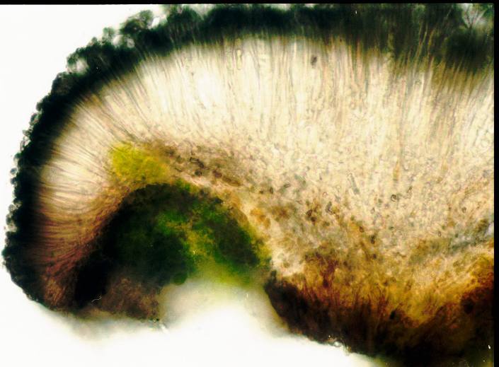
Lecidella stigmatea (Ach.) Hertel & Leuckert
Description: Photograph of a cross section of an apothecium of Lecidella stigmatea taken through a compound microscope (x400), showing a hyaline hypothecium. [Note: hypothecium of Lecidella carpathica is pale to midbrown.]Copyright: Ed Uebel
License: CC BY-SA 3.0
Identified by: E. C. Uebel
Date: 2006-08-01 00:00:00
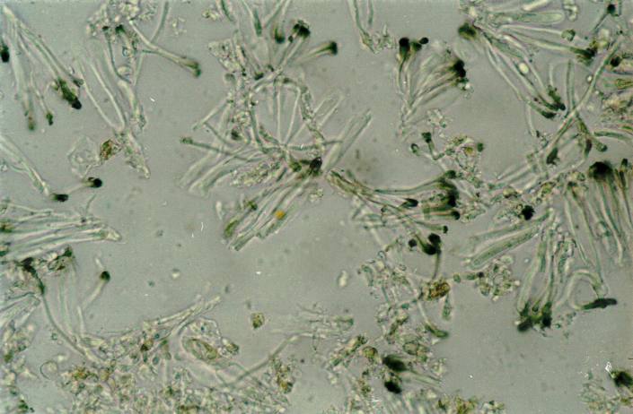
Lecidella stigmatea (Ach.) Hertel & Leuckert
Description: Photograph of a cross section of an apothecium of Lecidella stigmatea taken through a compound microscope (x1000), showing unbranched paraphyses that are easily separable in water.Copyright: Ed Uebel
License: CC BY-SA 3.0
Identified by: E. C. Uebel
Date: 2007-08-28 00:00:00

Porpidia crustulata (Ach.) Hertel & Knoph, 1984
Copyright: Ed UebelLicense: All Rights Reserved
Date: 0000-00-00 00:00:00

Porpidia crustulata (Ach.) Hertel & Knoph, 1984
Copyright: Ed UebelLicense: All Rights Reserved
Date: 0000-00-00 00:00:00

Porpidia crustulata (Ach.) Hertel & Knoph, 1984
Copyright: Ed UebelLicense: All Rights Reserved
Date: 0000-00-00 00:00:00

Porpidia crustulata (Ach.) Hertel & Knoph, 1984
Copyright: Ed UebelLicense: All Rights Reserved
Date: 0000-00-00 00:00:00

Porpidia crustulata (Ach.) Hertel & Knoph, 1984
Copyright: Ed UebelLicense: All Rights Reserved
Date: 0000-00-00 00:00:00

Porpidia crustulata (Ach.) Hertel & Knoph, 1984
Copyright: Ed UebelLicense: All Rights Reserved
Date: 0000-00-00 00:00:00

Porpidia crustulata (Ach.) Hertel & Knoph, 1984
Description: Photograph of an ascus of Porpidia crustulata stained with Lugol's solution (iodine solution) taken through a compound microscope (x1000) showing the staining of the tholus.Copyright: Ed Uebel
License: CC BY-SA 3.0
Identified by: E. C. Uebel
Date: 2005-09-04 00:00:00

Porpidia crustulata (Ach.) Hertel & Knoph, 1984
Description: Photograph of an ascus of Porpidia crustulata stained with Lugol's solution (iodine solution) taken through a compound microscope (x1000) showing the staining of the tholus.Copyright: Ed Uebel
License: CC BY-SA 3.0
Identified by: E. C. Uebel
Date: 2005-09-04 00:00:00

Porpidia crustulata (Ach.) Hertel & Knoph, 1984
Copyright: Ed UebelLicense: All Rights Reserved
Date: 0000-00-00 00:00:00

Porpidia crustulata (Ach.) Hertel & Knoph, 1984
Description: Photograph of a cross section of an apothecium from Porpidia crustulata taken through a compound microscope (x400) showing asci with 8 hyaline spores.Copyright: Ed Uebel
License: CC BY-SA 3.0
Identified by: E. C. Uebel
Date: 2005-09-04 00:00:00

Porpidia crustulata (Ach.) Hertel & Knoph, 1984
Copyright: James K. LindseyLicense: CC BY-SA 3.0
Date: 2007-02-28 00:00:00

Porpidia crustulata (Ach.) Hertel & Knoph, 1984
Copyright: James K. LindseyLicense: CC BY-SA 3.0
Date: 2007-02-28 00:00:00

Pseudocyphellaria aurata (Ach.) Vain.
Copyright: Paulo A. V. BorgesLicense: All Rights Reserved
Date: 2011-10-08 00:00:00

Circinaria calcarea (L.) A. Nordin, S. Savic & Tibell, 2010
Description: Photograph of a herbarium specimen, now in the Lichen Herbarium of the New York Botanical Garden.Copyright: Ed Uebel
License: CC BY-SA 3.0
Identified by: Richard E. Riefner
Date: 1981-07-21 00:00:00

Circinaria calcarea (L.) A. Nordin, S. Savic & Tibell, 2010
Description: Photograph of a herbarium specimen taken through a dissecting microscope, x6Copyright: Ed Uebel
License: CC BY-SA 3.0
Identified by: Richard E. Riefner
Date: 1981-07-21 00:00:00

Circinaria calcarea (L.) A. Nordin, S. Savic & Tibell, 2010
Description: Photograph of a herbarium specimen taken through a dissecting microscope, x40, specimen is now in the Lichen Herbarium of the New York Botanical Garden.Copyright: Ed Uebel
License: CC BY-SA 3.0
Identified by: Richard E. Riefner
Date: 1981-07-21 00:00:00

Circinaria calcarea (L.) A. Nordin, S. Savic & Tibell, 2010
Description: Photograph of a herbarium specimen taken through a dissecting microscope, x33, specimen is now in the Lichen Herbarium of the New York Botanical Garden.Copyright: Ed Uebel
License: CC BY-SA 3.0
Identified by: Richard E. Riefner
Date: 1981-07-21 00:00:00

Circinaria calcarea (L.) A. Nordin, S. Savic & Tibell, 2010
Description: Photograph of a herbarium specimen taken through a dissecting microscope (x21), specimen is now in the Lichen Herbarium of the New York Botanical Garden.Copyright: Ed Uebel
License: CC BY-SA 3.0
Identified by: Richard E. Riefner
Date: 1981-07-21 00:00:00

Mycoblastus sanguinarius (L.) Norman, 1853
Description: Photograph of a herbarium specimen.Copyright: Ed Uebel
License: CC BY-SA 3.0
Identified by: E. C. Uebel
Date: 2005-05-01 00:00:00

Mycoblastus sanguinarius (L.) Norman, 1853
Description: Photograph of a herbarium specimen.Copyright: Ed Uebel
License: CC BY-SA 3.0
Identified by: David H. S. Richardson
Date: 2005-05-01 00:00:00

Mycoblastus sanguinarius (L.) Norman, 1853
Description: Photograph of a herbarium specimen.Copyright: Ed Uebel
License: CC BY-SA 3.0
Identified by: David H. S. Richardson
Date: 2005-05-01 00:00:00

Mycoblastus sanguinarius (L.) Norman, 1853
Description: Photograph of a herbarium specimen taken through a dissecting microscope, x9.Copyright: Ed Uebel
License: CC BY-SA 3.0
Identified by: Irwin Brodo
Date: 2005-12-16 00:00:00

Mycoblastus sanguinarius (L.) Norman, 1853
Description: Photograph of a herbarium specimen taken through a dissecting microscope (x8) shows a bright red pigment in cut apothecia.Copyright: Ed Uebel
License: CC BY-SA 3.0
Identified by: Irwin Brodo
Date: 2005-12-16 00:00:00
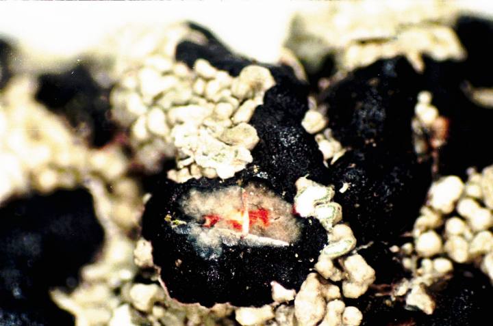
Mycoblastus sanguinarius (L.) Norman, 1853
Description: Photograph of a herbarium specimen taken through a dissecting microscope (x8) shows a bright red pigment in a cut apothecium.Copyright: Ed Uebel
License: CC BY-SA 3.0
Identified by: Irwin Brodo
Date: 2005-12-16 00:00:00
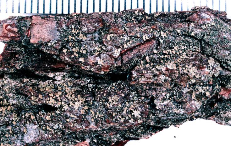
Hypocenomyce scalaris (Ach. ex Lilj.) M. Choisy, 1951
Description: Photograph of a herbarium specimen, now in the Lichen Herbarium of the New York Botanical Garden (millimeter scale).Copyright: Ed Uebel
License: CC BY-SA 3.0
Identified by: A. Norden & B. Ball
Date: 1974-02-16 00:00:00

Hypocenomyce scalaris (Ach. ex Lilj.) M. Choisy, 1951
Description: Photograph of a herbarium specimen, now in the Lichen Herbarium of the New York Botanical Garden (millimeter scale).Copyright: Ed Uebel
License: CC BY-SA 3.0
Identified by: A. Norden & B. Ball
Date: 1974-02-16 00:00:00

Hypocenomyce scalaris (Ach. ex Lilj.) M. Choisy, 1951
Description: Photograph of a herbarium specimen taken through a dissecting microscope (x40) showing squamules with powdery soredia on the underside of each squamule, specimen is now in the Lichen Herbarium of the New York Botanical Garden.Copyright: Ed Uebel
License: CC BY-SA 3.0
Identified by: Arnold W. Norden
Date: 1984-01-08 00:00:00

Hypocenomyce scalaris (Ach. ex Lilj.) M. Choisy, 1951
Description: Photograph of a herbarium specimen taken through a dissecting microscope (x40) showing squamules with powdery soredia on the underside of each squamule, specimen is now in the Lichen Herbarium of the New York Botanical Garden.Copyright: Ed Uebel
License: CC BY-SA 3.0
Identified by: Arnold W. Norden
Date: 1984-01-08 00:00:00

Hypocenomyce scalaris (Ach. ex Lilj.) M. Choisy, 1951
Description: Photograph of a herbarium specimen taken through a dissecting microscope (x40) showing squamules with powdery soredia on the underside of each squamule, specimen is now in the Lichen Herbarium of the New York Botanical Garden.Copyright: Ed Uebel
License: CC BY-SA 3.0
Identified by: Arnold W. Norden
Date: 1984-01-08 00:00:00

Cetraria aculeata (Schreb.) Fr.
Copyright: Alberto SalgueroLicense: CC BY-SA 3.0
Date: 2006-11-12 00:00:00

Cetraria aculeata (Schreb.) Fr.
Copyright: Alberto SalgueroLicense: CC BY-SA 3.0
Date: 0000-00-00 00:00:00

Cetrelia olivetorum (Nyl.) W. L. Culb. & C. F. Culb.
Description: Photograph of a herbarium specimen, now in the Lichen Herbarium of the New York Botanical Garden (millimeter scale).Copyright: Ed Uebel
License: CC BY-SA 3.0
Identified by: A. Norden & B. Norden
Date: 1974-06-07 00:00:00

Cetrelia olivetorum (Nyl.) W. L. Culb. & C. F. Culb.
Copyright: Ed UebelLicense: All Rights Reserved
Date: 0000-00-00 00:00:00

Cetrelia olivetorum (Nyl.) W. L. Culb. & C. F. Culb.
Description: Photograph of a herbarium specimen showing white pseudocyphellae, specimen is now in the Lichen Herbarium of the New York Botanical Garden.Copyright: Ed Uebel
License: CC BY-SA 3.0
Identified by: Elmer G. Worthley
Date: 1983-07-28 00:00:00

Cetrelia olivetorum (Nyl.) W. L. Culb. & C. F. Culb.
Description: Photograph of a herbarium specimen showing lobes with pseudocyphellae and marginal soredia. Chemical spot test on medulla: C+ red.Copyright: Ed Uebel
License: CC BY-SA 3.0
Identified by: E. C. Uebel
Date: 2005-04-15 00:00:00

Cetrelia olivetorum (Nyl.) W. L. Culb. & C. F. Culb.
Description: Photograph of a herbarium specimen showing lobes with a greenish-gray upper surface and a lower surface with brown edges and a black central portion. Chemical spot test on medulla: C+ red.Copyright: Ed Uebel
License: CC BY-SA 3.0
Identified by: E. C. Uebel
Date: 2005-04-15 00:00:00

Cetrelia olivetorum (Nyl.) W. L. Culb. & C. F. Culb.
Copyright: Ed UebelLicense: All Rights Reserved
Date: 0000-00-00 00:00:00

Cetrelia olivetorum (Nyl.) W. L. Culb. & C. F. Culb.
Copyright: Ed UebelLicense: All Rights Reserved
Date: 0000-00-00 00:00:00

Menegazzia terebrata (Hoffm.) A. Massal.
Copyright: OhWehLicense: CC-BY-SA-2.5
Date: 2008-05-03 00:00:00

Menegazzia terebrata (Hoffm.) A. Massal.
Copyright: OhWehLicense: CC-BY-SA-2.5
Date: 2008-05-03 00:00:00

Menegazzia terebrata (Hoffm.) A. Massal.
Copyright: OhWehLicense: CC-BY-SA-2.5
Date: 2008-05-03 00:00:00

Menegazzia terebrata (Hoffm.) A. Massal.
Copyright: Ed UebelLicense: All Rights Reserved
Date: 0000-00-00 00:00:00

Menegazzia terebrata (Hoffm.) A. Massal.
Copyright: Ed UebelLicense: All Rights Reserved
Date: 0000-00-00 00:00:00

Menegazzia terebrata (Hoffm.) A. Massal.
Copyright: Ed UebelLicense: All Rights Reserved
Date: 0000-00-00 00:00:00

Parmotrema reticulatum (Taylor) M. Choisy
Description: Photograph of a herbarium specimen, now in the Lichen Herbarium of the New York Botanical Garden.Copyright: Ed Uebel
License: CC BY-SA 3.0
Identified by: Elmer G. Worthley
Date: 0000-00-00 00:00:00

Parmotrema reticulatum (Taylor) M. Choisy
Description: Photograph of a herbarium specimen showing reticulate cracks in the upper surface of the lobes, specimen is now in the Lichen Herbarium of the New York Botanical Garden.Copyright: Ed Uebel
License: CC BY-SA 3.0
Identified by: Elmer G. Worthley
Date: 0000-00-00 00:00:00

Parmotrema reticulatum (Taylor) M. Choisy
Description: Photograph of a herbarium specimen, now in the Lichen Herbarium of the New York Botanical Garden.Copyright: Ed Uebel
License: CC BY-SA 3.0
Identified by: Elmer G. Worthley
Date: 1978-03-14 00:00:00

Parmotrema reticulatum (Taylor) M. Choisy
Description: Photograph of a herbarium specimen showing reticulate cracks in the upper surface of the lobes, specimen is now in the Lichen Herbarium of the New York Botanical Garden.Copyright: Ed Uebel
License: CC BY-SA 3.0
Identified by: Elmer G. Worthley, A. Norden & A. Skorepa
Date: 1977-05-07 00:00:00

Parmotrema reticulatum (Taylor) M. Choisy
Description: Chemistry: Medulla- PD+ orange, K+ yellow turning red, C- Photograph of a herbarium specimen showing fine reticulate cracking in the upper surface of a lobe.Copyright: Ed Uebel
License: CC BY-SA 3.0
Identified by: E. C. Uebel
Date: 2005-04-23 00:00:00

Usnea krogiana P. Clerc
Copyright: Paulo Henrique SilvaLicense: All Rights Reserved
Date: 2009-05-20 00:00:00

Usnea krogiana P. Clerc
Copyright: Paulo Henrique SilvaLicense: All Rights Reserved
Date: 2009-05-20 00:00:00

Usnea krogiana P. Clerc
Copyright: Paulo Henrique SilvaLicense: All Rights Reserved
Date: 2009-07-09 00:00:00

Peltigera hymenina (Ach.) Delise, 1830
Copyright: Bernd HaynoldLicense: CC BY-SA 3.0
Date: 2008-02-28 00:00:00

Peltigera hymenina (Ach.) Delise, 1830
Copyright: Bernd HaynoldLicense: CC BY-SA 3.0
Date: 2008-02-28 00:00:00

Peltigera melanorrhiza Purvis, P. James & Vitik.
Copyright: Paulo Henrique SilvaLicense: All Rights Reserved
Date: 0000-00-00 00:00:00

Peltigera melanorrhiza Purvis, P. James & Vitik.
Copyright: Paulo Henrique SilvaLicense: All Rights Reserved
Date: 0000-00-00 00:00:00

Peltigera melanorrhiza Purvis, P. James & Vitik.
Copyright: Paulo Henrique SilvaLicense: All Rights Reserved
Date: 0000-00-00 00:00:00

Peltigera melanorrhiza Purvis, P. James & Vitik.
Copyright: Paulo Henrique SilvaLicense: All Rights Reserved
Date: 0000-00-00 00:00:00

Lepra amara (Ach.) Hafellner, 2016
Copyright: James K. LindseyLicense: CC BY-SA 3.0
Date: 2007-03-02 00:00:00

Lepra amara (Ach.) Hafellner, 2016
Copyright: Kristian PetersLicense: CC BY-SA 3.0
Date: 2006-11-10 00:00:00

Lepra amara (Ach.) Hafellner, 2016
Description: Photograph of a herbarium specimen showing sorediate fruiting warts.Copyright: Ed Uebel
License: CC BY-SA 3.0
Identified by: David H. S. Richardson
Date: 2005-05-01 00:00:00

Lepra amara (Ach.) Hafellner, 2016
Copyright: James K. LindseyLicense: CC BY-SA 3.0
Date: 2007-03-02 00:00:00

Lepra amara (Ach.) Hafellner, 2016
Description: Photograph of a herbarium specimen showing sorediate fruiting warts.Copyright: Ed Uebel
License: CC BY-SA 3.0
Identified by: David H. S. Richardson
Date: 2005-05-01 00:00:00

Amandinea punctata (Hoffm.) Coppins & Scheid., 1993
Description: Photograph of a herbarium specimen taken through a dissecting microscope showing a barely perceptible thallus and black marginless apothecia measuring ca. 0.3 mm in diameter.Copyright: Ed Uebel
License: CC BY-SA 3.0
Identified by: E. C. Uebel
Date: 2003-08-26 00:00:00

Amandinea punctata (Hoffm.) Coppins & Scheid., 1993
Copyright: James K. LindseyLicense: CC BY-SA 3.0
Date: 2007-02-27 00:00:00
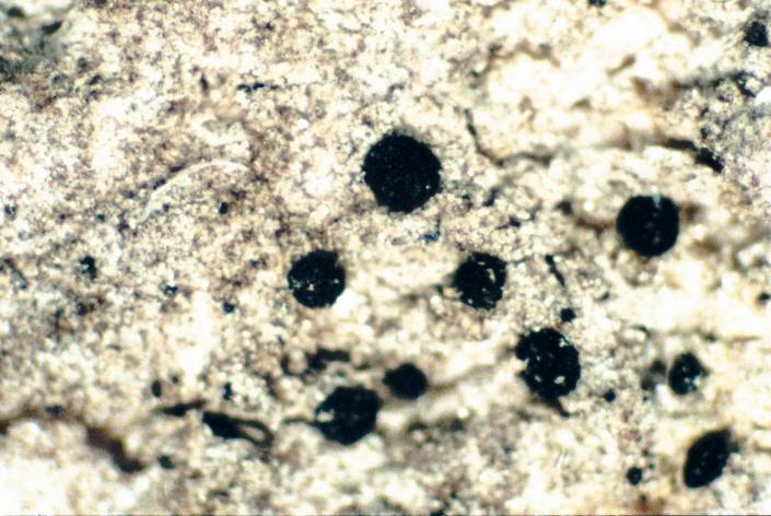
Amandinea punctata (Hoffm.) Coppins & Scheid., 1993
Description: Photograph of a herbarium specimen taken through a dissecting microscope showing a barely perceptible thallus and black marginless apothecia measuring ca. 0.3 mm in diameter.Copyright: Ed Uebel
License: CC BY-SA 3.0
Identified by: E. C. Uebel
Date: 2003-08-26 00:00:00

Amandinea punctata (Hoffm.) Coppins & Scheid., 1993
Copyright: James K. LindseyLicense: CC BY-SA 3.0
Date: 2007-02-27 00:00:00

Amandinea punctata (Hoffm.) Coppins & Scheid., 1993
Description: Photograph of a herbarium specimen taken through a dissecting microscope showing a barely perceptible thallus and black marginless apothecia measuring ca. 0.3 mm in diameter.Copyright: Ed Uebel
License: CC BY-SA 3.0
Identified by: E. C. Uebel
Date: 2003-08-26 00:00:00

Amandinea punctata (Hoffm.) Coppins & Scheid., 1993
Description: Photograph of a cross section of an apothecium from Amandinea punctata taken through a compound microscope, x400. The exciple is uniformly pigmented dark brown; the epihymenium is brown; the hypothecium is brown black.Copyright: Ed Uebel
License: CC BY-SA 3.0
Identified by: E. C. Uebel
Date: 2003-08-26 00:00:00

Amandinea punctata (Hoffm.) Coppins & Scheid., 1993
Description: Photograph of a cross section of an apothecium of Amandinea punctata taken through a compound microscope (x1000), showing 8 brown 1-septate spores per ascus.Copyright: Ed Uebel
License: CC BY-SA 3.0
Identified by: E. C. Uebel
Date: 2003-08-26 00:00:00

Amandinea punctata (Hoffm.) Coppins & Scheid., 1993
Description: Photograph of a brown, 1-septate spore from an apothecium of Amandinea punctata taken through a compound microscope, x1000 (spore measures 8 x 3 microns).Copyright: Ed Uebel
License: CC BY-SA 3.0
Identified by: E. C. Uebel
Date: 2003-08-26 00:00:00

Physcia caesia (Hoffm.) Hampe ex Fürnr., 1839
Description: Photograph of a herbarium specimen, now in the Lichen Herbarium of the New York Botanical Garden (millimeter scale).Copyright: Ed Uebel
License: CC BY-SA 3.0
Identified by: Richard E. Riefner
Date: 1981-06-27 00:00:00

Physcia caesia (Hoffm.) Hampe ex Fürnr., 1839
Description: Photograph of a herbarium specimen, now in the Lichen Herbarium of the New York Botanical Garden (millimeter scale).Copyright: Ed Uebel
License: CC BY-SA 3.0
Identified by: Richard E. Riefner
Date: 1981-06-27 00:00:00
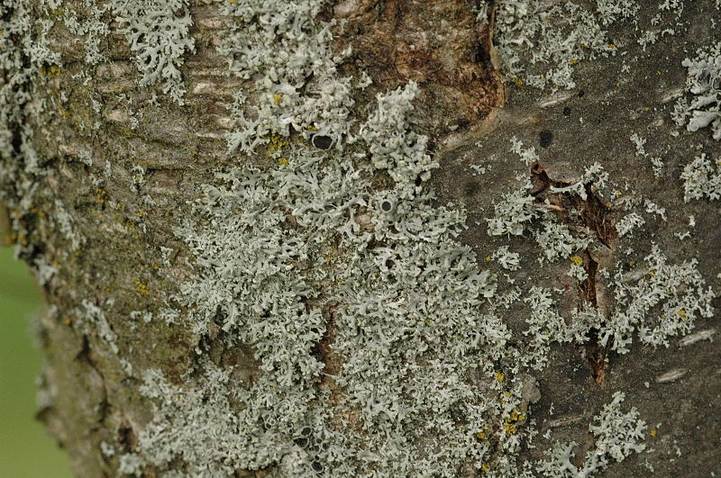
Physcia stellaris (L.) Nyl., 1856
Copyright: James K. LindseyLicense: CC BY-SA 3.0
Date: 2004-09-26 00:00:00
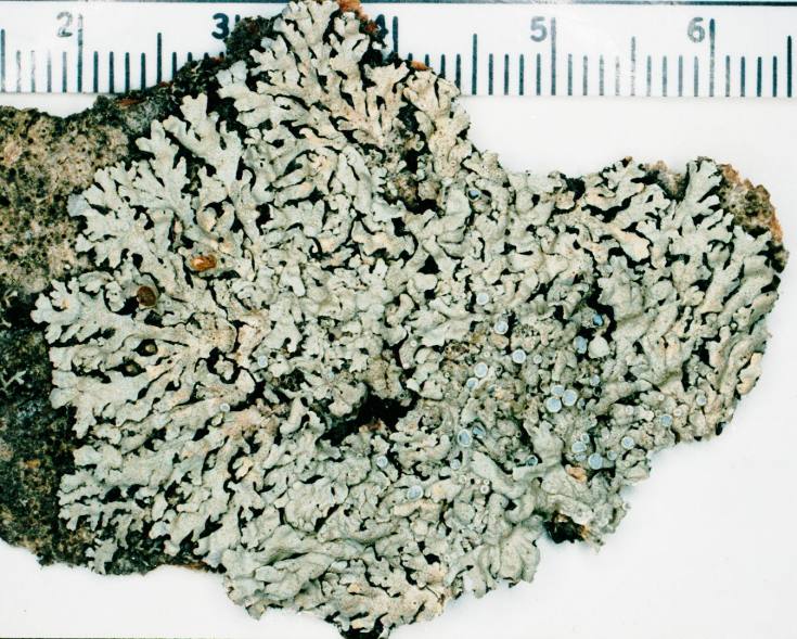
Physcia stellaris (L.) Nyl., 1856
Description: Photograph of a herbarium specimen, now in the Lichen Herbarium of the New York Botanical Garden.Copyright: Ed Uebel
License: CC BY-SA 3.0
Identified by: Richard E. Riefner
Date: 1981-07-07 00:00:00
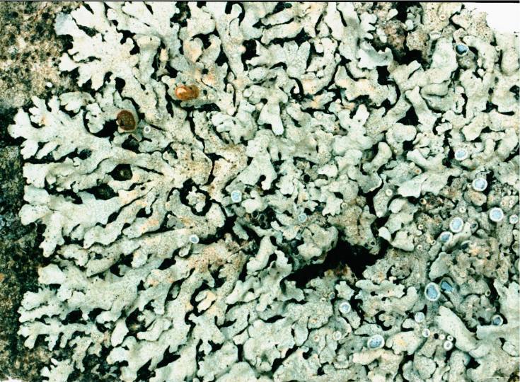
Physcia stellaris (L.) Nyl., 1856
Description: Photograph of a herbarium specimen, now in the Lichen Herbarium of the New York Botanical Garden.Copyright: Ed Uebel
License: CC BY-SA 3.0
Identified by: Richard E. Riefner
Date: 1981-07-07 00:00:00
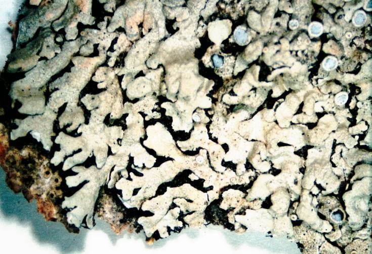
Physcia stellaris (L.) Nyl., 1856
Description: Photograph of a herbarium specimen, now in the Lichen Herbarium of the New York Botanical Garden.Copyright: Ed Uebel
License: CC BY-SA 3.0
Identified by: Richard E. Riefner
Date: 1981-07-07 00:00:00
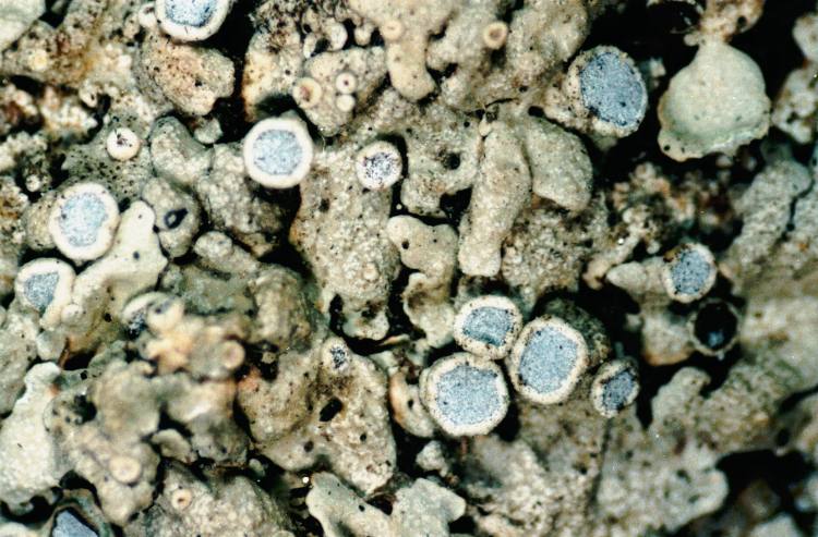
Physcia stellaris (L.) Nyl., 1856
Description: Photograph of a herbarium specimen, now in the Lichen Herbarium of the New York Botanical Garden.Copyright: Ed Uebel
License: CC BY-SA 3.0
Identified by: Richard E. Riefner
Date: 1981-07-07 00:00:00
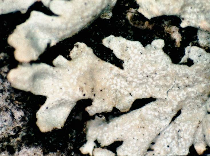
Physcia stellaris (L.) Nyl., 1856
Description: Photograph of a herbarium specimen showing close-up of lobes, specimen is now in the Lichen Herbarium of the New York Botanical Garden.Copyright: Ed Uebel
License: CC BY-SA 3.0
Identified by: Richard E. Riefner
Date: 1981-07-07 00:00:00
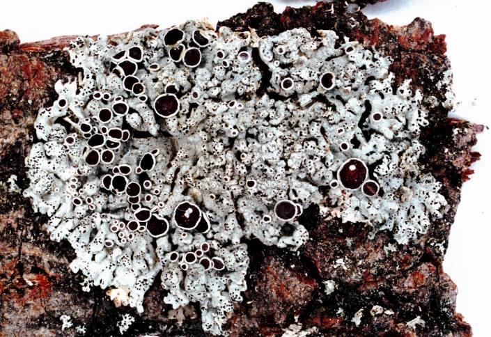
Physcia stellaris (L.) Nyl., 1856
Description: Photograph of a herbarium specimen.Copyright: Ed Uebel
License: CC BY-SA 3.0
Identified by: E. C. Uebel
Date: 2005-05-01 00:00:00
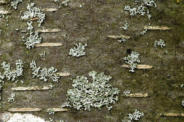
Physcia tenella (Scop.) DC., 1805
Copyright: James K. LindseyLicense: CC BY-SA 3.0
Date: 2004-09-26 00:00:00
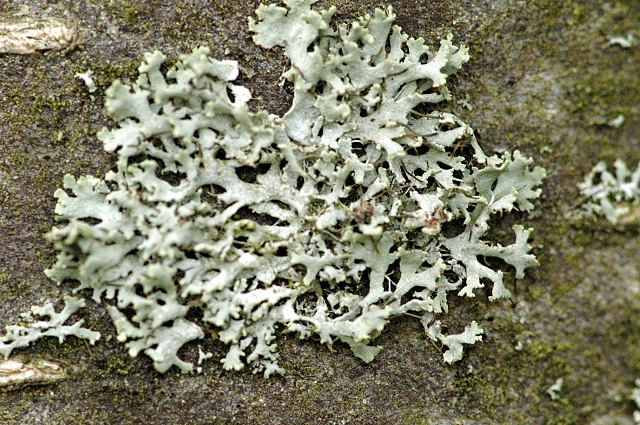
Physcia tenella (Scop.) DC., 1805
Copyright: James K. LindseyLicense: CC BY-SA 3.0
Date: 2004-09-26 00:00:00

Ramalina azorica Aptroot & Schumm
Copyright: Paulo Henrique SilvaLicense: All Rights Reserved
Date: 2010-07-14 00:00:00

Ramalina azorica Aptroot & Schumm
Copyright: Paulo Henrique SilvaLicense: All Rights Reserved
Date: 2010-07-14 00:00:00

Ramalina azorica Aptroot & Schumm
Copyright: Paulo Henrique SilvaLicense: All Rights Reserved
Date: 2010-07-14 00:00:00

Ramalina azorica Aptroot & Schumm
Copyright: Paulo Henrique SilvaLicense: All Rights Reserved
Date: 2010-07-14 00:00:00

Ramalina azorica Aptroot & Schumm
Copyright: Paulo Henrique SilvaLicense: All Rights Reserved
Date: 2010-07-14 00:00:00

Rhizocarpon hochstetteri (Körb.) Vain., 1922
Description: Photograph of a herbarium specimen on a small stone. Photographed through a dissecting microscope, x 6.Copyright: Ed Uebel
License: CC BY-SA 3.0
Identified by: E. C. Uebel
Date: 2003-07-16 00:00:00
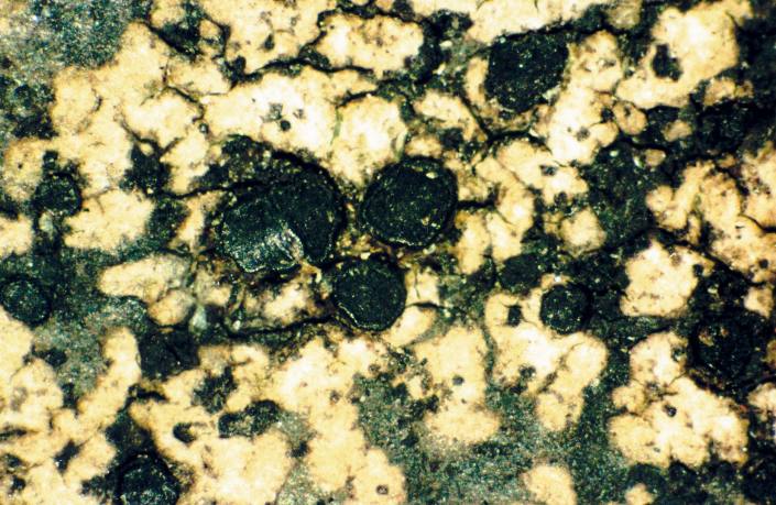
Rhizocarpon hochstetteri (Körb.) Vain., 1922
Description: Photograph of a herbarium specimen on a small stone. Photographed through a dissecting microscope, x 40.Copyright: Ed Uebel
License: CC BY-SA 3.0
Identified by: E. C. Uebel
Date: 2003-07-16 00:00:00

Rhizocarpon hochstetteri (Körb.) Vain., 1922
Description: Photograph of a herbarium specimen on a small stone. Photographed through a dissecting microscope, x 40.Copyright: Ed Uebel
License: CC BY-SA 3.0
Identified by: E. C. Uebel
Date: 2003-07-16 00:00:00

Rhizocarpon hochstetteri (Körb.) Vain., 1922
Description: Photograph of a herbarium specimen on a small stone. Photographed through a dissecting microscope, x 40.Copyright: Ed Uebel
License: CC BY-SA 3.0
Identified by: E. C. Uebel
Date: 2003-07-16 00:00:00

Rhizocarpon hochstetteri (Körb.) Vain., 1922
Description: Photograph of an ascus from an apothecium of Rhizocarpon hochstetteri taken through a compound microscope (x1000), showing 8, 1-septate spores per ascus.Copyright: Ed Uebel
License: CC BY-SA 3.0
Identified by: E. C. Uebel
Date: 2003-07-16 00:00:00

Rhizocarpon hochstetteri (Körb.) Vain., 1922
Description: Photograph of 1-septate spores from an apothecium of Rhizocarpon hochstetteri taken through a compound microscope, x1000. Spores measure ca. 18 x 9 microns.Copyright: Ed Uebel
License: CC BY-SA 3.0
Identified by: E. C. Uebel
Date: 2003-07-16 00:00:00

Rhizocarpon hochstetteri (Körb.) Vain., 1922
Description: Photograph of 1-septate spores from an apothecium of Rhizocarpon hochstetteri taken through a compound microscope, x1000.Copyright: Ed Uebel
License: CC BY-SA 3.0
Identified by: E. C. Uebel
Date: 2003-07-16 00:00:00

Rhizocarpon hochstetteri (Körb.) Vain., 1922
Description: Photograph of a 1-septate spore from an apothecium of Rhizocarpon hochstetteri taken through a compound microscope, x1000. Spore length 20-25 microns.Copyright: Ed Uebel
License: CC BY-SA 3.0
Identified by: E. C. Uebel
Date: 2003-07-16 00:00:00

Loxospora elatina (Ach.) A. Massal., 1852
Description: Photograph of a herbarium specimen.Copyright: Ed Uebel
License: CC BY-SA 3.0
Identified by: David H. S. Richardson
Date: 2005-05-01 00:00:00

Loxospora elatina (Ach.) A. Massal., 1852
Description: Photograph of a herbarium specimen.Copyright: Ed Uebel
License: CC BY-SA 3.0
Identified by: David H. S. Richardson
Date: 2005-05-01 00:00:00

Loxospora elatina (Ach.) A. Massal., 1852
Description: Photograph of a herbarium specimen taken through a dissecting microscope, x40.Copyright: Ed Uebel
License: CC BY-SA 3.0
Identified by: David H. S. Richardson
Date: 2005-05-01 00:00:00

Loxospora elatina (Ach.) A. Massal., 1852
Description: Photograph of a herbarium specimen taken through a dissecting microscope, x15.Copyright: Ed Uebel
License: CC BY-SA 3.0
Identified by: David H. S. Richardson
Date: 2005-05-01 00:00:00

Loxospora elatina (Ach.) A. Massal., 1852
Description: Photograph of a herbarium specimen taken through a dissecting microscope (x40), showing apothecia.Copyright: Ed Uebel
License: CC BY-SA 3.0
Identified by: David H. S. Richardson
Date: 2005-05-01 00:00:00
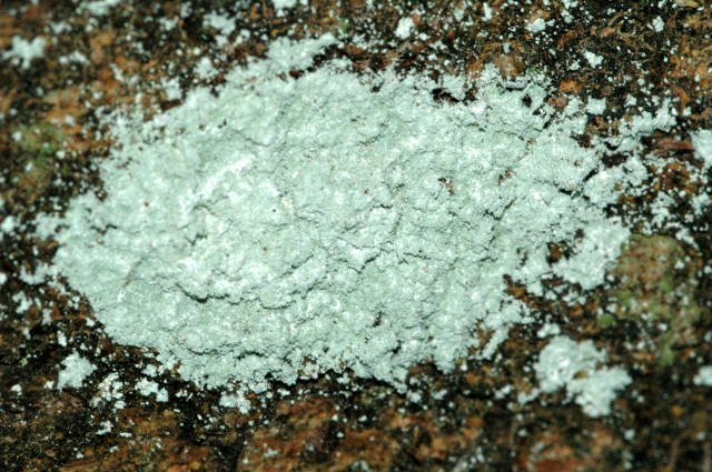
Lepraria incana (L.) Ach.
Copyright: James K. LindseyLicense: CC BY-SA 3.0
Date: 2004-09-04 00:00:00
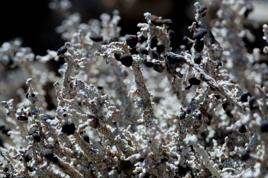
Stereocaulon azoreum (Schaer.) Nyl.
Copyright: Paulo Henrique SilvaLicense: All Rights Reserved
Date: 2010-07-14 00:00:00
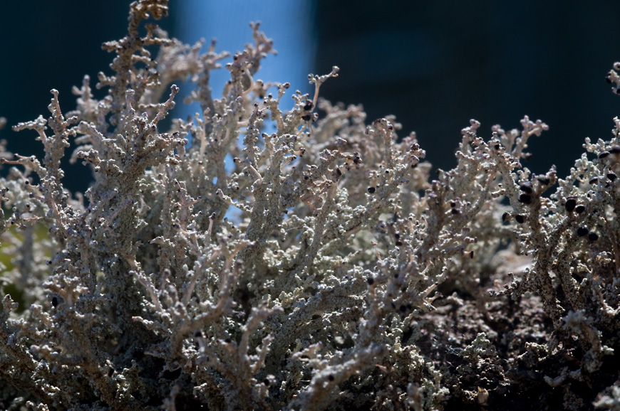
Stereocaulon azoreum (Schaer.) Nyl.
Copyright: Paulo Henrique SilvaLicense: All Rights Reserved
Date: 2010-07-14 00:00:00
Stereocaulon azoreum (Schaer.) Nyl.
Copyright: Paulo Henrique SilvaLicense: All Rights Reserved
Date: 2010-07-14 00:00:00
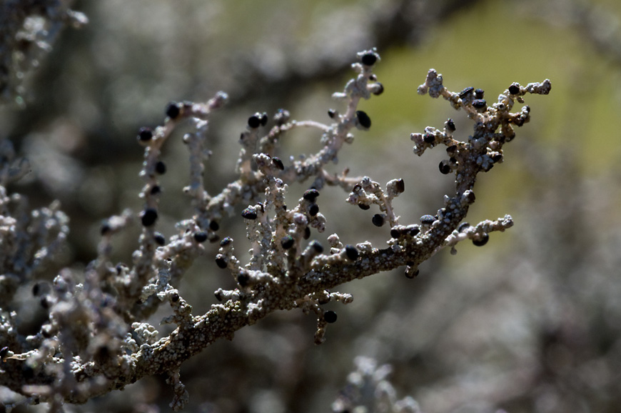
Stereocaulon azoreum (Schaer.) Nyl.
Copyright: Paulo Henrique SilvaLicense: All Rights Reserved
Date: 2010-07-14 00:00:00

Flavoplaca citrina (Hoffmann) Arup, Frödén & Søchting, 2013
Copyright: Ed UebelLicense: CC BY-SA 3.0
Date: 2002-03-11 00:00:00

Flavoplaca citrina (Hoffmann) Arup, Frödén & Søchting, 2013
Description: Photograph of a herbarium specimen taken through a dissecting microscope (x7) showing a yellow sorediate thallus, specimen is now in the Lichen Herbarium of the New York Botanical Garden.Copyright: Ed Uebel
License: CC BY-SA 3.0
Identified by: Elmer G. Worthley
Date: 1980-03-30 00:00:00

Flavoplaca citrina (Hoffmann) Arup, Frödén & Søchting, 2013
Description: Photograph of a herbarium specimen taken through a dissecting microscope (x7) showing a yellow sorediate thallus), specimen is now in the Lichen Herbarium of the New York Botanical Garden.Copyright: Ed Uebel
License: CC BY-SA 3.0
Identified by: Elmer G. Worthley
Date: 1980-03-30 00:00:00

Flavoplaca citrina (Hoffmann) Arup, Frödén & Søchting, 2013
Description: Close-up view of Caloplaca citrina growing the wall of a concrete bridge.Copyright: Ed Uebel
License: CC BY-SA 3.0
Date: 2001-06-25 00:00:00

Flavoplaca citrina (Hoffmann) Arup, Frödén & Søchting, 2013
Description: Close-up view of Caloplaca citrina growing the wall of a concrete bridge.Copyright: Ed Uebel
License: CC BY-SA 3.0
Date: 2001-06-25 00:00:00

Flavoplaca citrina (Hoffmann) Arup, Frödén & Søchting, 2013
Description: Close-up view of an apothecium of Caloplaca citrina from thallus scaped from a concrete wall; photographed through a dissecting microscope, x 40.Copyright: Ed Uebel
License: CC BY-SA 3.0
Date: 2001-06-25 00:00:00

Flavoplaca citrina (Hoffmann) Arup, Frödén & Søchting, 2013
Description: Yellow stainCopyright: Ed Uebel
License: CC BY-SA 3.0
Date: 2001-06-25 00:00:00

Flavoplaca citrina (Hoffmann) Arup, Frödén & Søchting, 2013
Copyright: Ed UebelLicense: CC BY-SA 3.0
Date: 2001-06-25 00:00:00

Flavoplaca citrina (Hoffmann) Arup, Frödén & Søchting, 2013
Copyright: Ed UebelLicense: CC BY-SA 3.0
Date: 2002-03-11 00:00:00

Gyalolechia flavorubescens (Huds.) Søchting, Frödén & Arup, 2013
Description: Photograph of a herbarium specimen taken through a dissecting microscope (x40) showing apothecia with dark orange disks and pale orange margins, specimen is now in the Lichen Herbarium of the New York Botanical Garden.Copyright: Ed Uebel
License: CC BY-SA 3.0
Identified by: Elmer G. Worthley
Date: 1977-04-30 00:00:00

Gyalolechia flavorubescens (Huds.) Søchting, Frödén & Arup, 2013
Description: Photograph of a herbarium specimen taken through a dissecting microscope (x40) showing apothecia with dark orange disks and pale orange margins, specimen is now in the Lichen Herbarium of the New York Botanical Garden.Copyright: Ed Uebel
License: CC BY-SA 3.0
Identified by: Elmer G. Worthley
Date: 1977-04-30 00:00:00

Gyalolechia flavovirescens (Wulfen) Søchting, Frödén & Arup, 2013
Description: Photograph of a herbarium specimen taken through a dissecting microscope (x6) showing a sulphur-yellow thallus and apothecia with orange disks and yellow margins, specimen is now in the Lichen Herbarium of the New York Botanical Garden.Copyright: Ed Uebel
License: CC BY-SA 3.0
Identified by: Richard E. Riefner
Date: 1981-07-23 00:00:00

Gyalolechia flavovirescens (Wulfen) Søchting, Frödén & Arup, 2013
Description: Photograph of a herbarium specimen taken through a dissecting microscope (x40) showing a sulphur-yellow thallus and apothecia with orange disks and yellow margins, specimen is now in the Lichen Herbarium of the New York Botanical Garden.Copyright: Ed Uebel
License: CC BY-SA 3.0
Identified by: Richard E. Riefner
Date: 1981-07-23 00:00:00

Gyalolechia flavovirescens (Wulfen) Søchting, Frödén & Arup, 2013
Description: Photograph of a herbarium specimen taken through a dissecting microscope (x40) showing a sulphur-yellow thallus and apothecia with orange disks and yellow margins, specimen is now in the Lichen Herbarium of the New York Botanical Garden.Copyright: Ed Uebel
License: CC BY-SA 3.0
Identified by: Richard E. Riefner
Date: 1981-07-23 00:00:00

Cerothallia luteoalba (Turner) Arup, Frödén & Søchting, 2013
Description: (photograph of a herbarium specimen) This specimen was in Dr. Elmer G. Worthley's personal lichen herbarium and the label on the packet reads as follows: "Lichenes suecici/ Caloplaca luteoalba (Turn.) Närke/ Par. Ringkarleby: Asplunda/ VI_VIII 1899/ leg.Copyright: Ed Uebel
License: CC BY-SA 3.0
Date: 0000-00-00 00:00:00

Cerothallia luteoalba (Turner) Arup, Frödén & Søchting, 2013
Description: (photograph of a herbarium specimen taken through a dissecting microscope (x6) showing thallus and orange apothecia) This specimen was in Dr. Elmer G. Worthley's personal lichen herbarium and the label on the packet reads as follows: "Lichenes suecici/ CaCopyright: Ed Uebel
License: CC BY-SA 3.0
Date: 0000-00-00 00:00:00

Cerothallia luteoalba (Turner) Arup, Frödén & Søchting, 2013
Description: (photograph of a herbarium specimen taken through a dissecting microscope (x17) showing thallus and orange apothecia) This specimen was in Dr. Elmer G. Worthley's personal lichen herbarium and the label on the packet reads as follows: "Lichenes suecici/ CCopyright: Ed Uebel
License: CC BY-SA 3.0
Date: 0000-00-00 00:00:00

Cerothallia luteoalba (Turner) Arup, Frödén & Søchting, 2013
Description: (photograph of a herbarium specimen taken through a dissecting microscope (x40) showing thallus and orange apothecia) This specimen was in Dr. Elmer G. Worthley's personal lichen herbarium and the label on the packet reads as follows: "Lichenes suecici/ CCopyright: Ed Uebel
License: CC BY-SA 3.0
Date: 0000-00-00 00:00:00

Cerothallia luteoalba (Turner) Arup, Frödén & Søchting, 2013
Description: (photograph of a herbarium specimen taken through a dissecting microscope (x40) showing thallus and orange apothecia) This specimen was in Dr. Elmer G. Worthley's personal lichen herbarium and the label on the packet reads as follows: "Lichenes suecici/ CCopyright: Ed Uebel
License: CC BY-SA 3.0
Date: 0000-00-00 00:00:00

Calogaya saxicola (Hoffm.) Vondrák, 2016
Copyright: James K. LindseyLicense: CC BY-SA 3.0
Date: 2004-10-14 00:00:00

Calogaya saxicola (Hoffm.) Vondrák, 2016
Copyright: James K. LindseyLicense: CC BY-SA 3.0
Date: 2004-10-14 00:00:00

Gyalolechia fulgens (Swartz) Søchting, Frödén & Arup, 2013
Copyright: Bernd HaynoldLicense: CC BY-SA 3.0
Date: 2006-05-14 00:00:00

Gyalolechia fulgens (Swartz) Søchting, Frödén & Arup, 2013
Copyright: Bernd HaynoldLicense: CC BY-SA 3.0
Date: 2006-05-14 00:00:00

Gyalolechia fulgens (Swartz) Søchting, Frödén & Arup, 2013
Description: Fulgensia fulgens, above, yellow Psora decipiens, below, redCopyright: Bernd Haynold
License: CC BY-SA 3.0
Date: 2007-10-14 00:00:00

Polycauliona candelaria (Linnaeus) Frödén, Arup & Søchting, 2013
Copyright: James K. LindseyLicense: CC BY-SA 3.0
Date: 2005-01-10 00:00:00

Polycauliona candelaria (Linnaeus) Frödén, Arup & Søchting, 2013
Copyright: James K. LindseyLicense: CC BY-SA 3.0
Date: 2005-01-10 00:00:00

Xanthoria elegans (Link) Th. Fr.
Copyright: AnsgarLicense: All Rights Reserved
Date: 0000-00-00 00:00:00

Xanthoria elegans (Link) Th. Fr.
Copyright: Ed UebelLicense: All Rights Reserved
Date: 0000-00-00 00:00:00

Xanthoria elegans (Link) Th. Fr.
Copyright: TigerenteLicense: All Rights Reserved
Date: 0000-00-00 00:00:00

Xanthoria elegans (Link) Th. Fr.
Copyright: Ed UebelLicense: All Rights Reserved
Date: 0000-00-00 00:00:00

Xanthoria elegans (Link) Th. Fr.
Copyright: TigerenteLicense: CC BY-SA 3.0
Date: 2006-07-02 00:00:00

Xanthoria elegans (Link) Th. Fr.
Description: Photograph of a herbarium specimen taken through a dissecting microscope (x40) showing convex, reddish-orange lobes, specimen is now in the Lichen Herbarium of the New York Botanical Garden.Copyright: Ed Uebel
License: CC BY-SA 3.0
Date: 1981-06-27 00:00:00

Xanthoria elegans (Link) Th. Fr.
Copyright: Ed UebelLicense: All Rights Reserved
Date: 0000-00-00 00:00:00

Xanthomendoza fallax (Arnold) Søchting, Kärnefelt & S.Y. Kondr., 2002
Description: Photograph of a herbarium specimen, now in the Lichen Herbarium of the New York Botanical Garden.Copyright: Ed Uebel
License: CC BY-SA 3.0
Identified by: Elmer G. Worthley
Date: 0000-00-00 00:00:00

Xanthomendoza fallax (Arnold) Søchting, Kärnefelt & S.Y. Kondr., 2002
Description: Photograph of a herbarium specimen taken through a dissecting microscope (x6) showing labriform soralia on the lobe tips, specimen is now in the Lichen Herbarium of the New York Botanical Garden.Copyright: Ed Uebel
License: CC BY-SA 3.0
Identified by: Elmer G. Worthley
Date: 0000-00-00 00:00:00

Xanthomendoza fallax (Arnold) Søchting, Kärnefelt & S.Y. Kondr., 2002
Description: Photograph of a herbarium specimen taken through a dissecting microscope (x40) showing labriform soralia on the lobe tips, specimen is now in the Lichen Herbarium of the New York Botanical Garden.Copyright: Ed Uebel
License: CC BY-SA 3.0
Identified by: Elmer G. Worthley
Date: 0000-00-00 00:00:00

Xanthomendoza fallax (Arnold) Søchting, Kärnefelt & S.Y. Kondr., 2002
Description: Photograph of a herbarium specimen taken through a dissecting microscope (x40) showing labriform soralia on the lobe tips, specimen is now in the Lichen Herbarium of the New York Botanical Garden.Copyright: Ed Uebel
License: CC BY-SA 3.0
Identified by: Elmer G. Worthley
Date: 0000-00-00 00:00:00

Xanthomendoza fallax (Arnold) Søchting, Kärnefelt & S.Y. Kondr., 2002
Description: Photograph of a herbarium specimen taken through a dissecting microscope (x40) showing labriform soralia on the lobe tips, specimen is now in the Lichen Herbarium of the New York Botanical Garden.Copyright: Ed Uebel
License: CC BY-SA 3.0
Identified by: Elmer G. Worthley
Date: 0000-00-00 00:00:00

Diploschistes muscorum (Scop.) R. Sant., 1980
Copyright: Ed UebelLicense: All Rights Reserved
Date: 0000-00-00 00:00:00

Diploschistes muscorum (Scop.) R. Sant., 1980
Description: Photograph of a herbarium specimen taken through a dissecting microscope, x40.Copyright: Ed Uebel
License: CC BY-SA 3.0
Identified by: E. C. Uebel
Date: 2005-05-02 00:00:00

Diploschistes muscorum (Scop.) R. Sant., 1980
Description: Photograph of a herbarium specimen taken through a dissecting microscope (x18) showing apothecia with relatively deep, wide cavities.Copyright: Ed Uebel
License: CC BY-SA 3.0
Identified by: E. C. Uebel
Date: 2005-05-02 00:00:00

Diploschistes muscorum (Scop.) R. Sant., 1980
Description: Photograph of a herbarium specimen taken through a dissecting microscope (x40) showing apothecia with relatively deep, wide cavities.Copyright: Ed Uebel
License: CC BY-SA 3.0
Identified by: E. C. Uebel
Date: 2005-05-02 00:00:00

Diploschistes muscorum (Scop.) R. Sant., 1980
Description: Photograph of a cross section of an apothecium of D. muscorum taken through a compound microscope (x400) showing 4 muriform spores per ascus.Copyright: Ed Uebel
License: CC BY-SA 3.0
Identified by: E. C. Uebel
Date: 2003-08-24 00:00:00

Diploschistes muscorum (Scop.) R. Sant., 1980
Description: Photograph of a muriform spore from D. muscorum taken through a compound microscope, x1000. (spore measures 30 x 10 microns)Copyright: Ed Uebel
License: CC BY-SA 3.0
Identified by: E. C. Uebel
Date: 2003-08-24 00:00:00